
Fig. 3
- ID
- ZDB-IMAGE-190925-10
- Publication
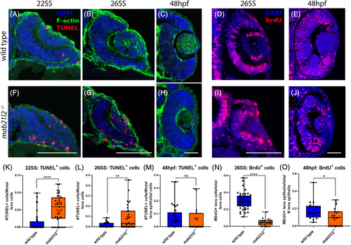
- Gath et al., 2019 - Zebrafish mab21l2 mutants possess severe defects in optic cup morphogenesis, lens and cornea development
- All Figures
- Figures for Gath et al., 2019
|
Fig. 3