
Fig. 5
- ID
- ZDB-IMAGE-190916-4
- Publication
- Toms et al., 2019 - Phagosomal and mitochondrial alterations in RPE may contribute to KCNJ13 retinopathy
- All Figures
- Figures for Toms et al., 2019
|
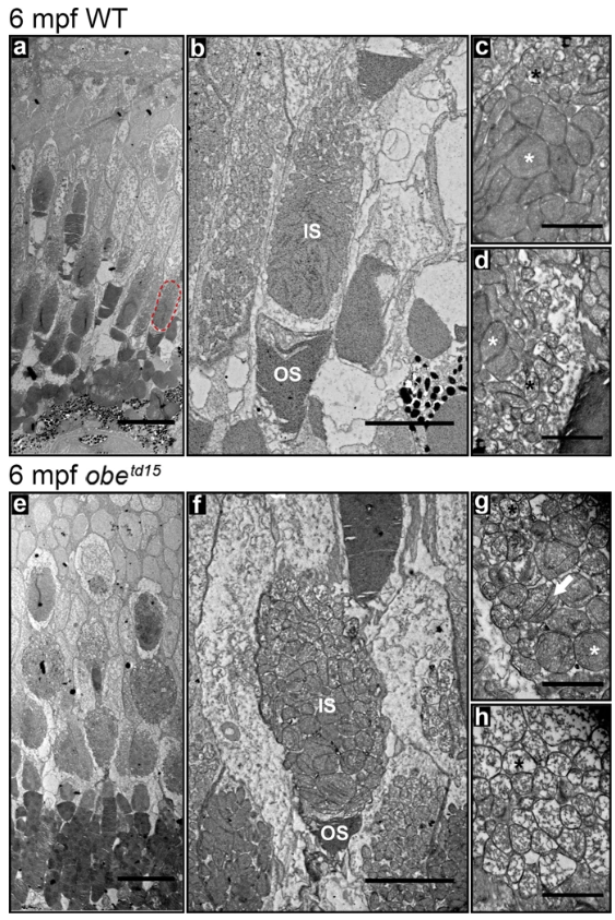
Fig. 5
Altered mitochondrial morphology in the obetd15 cone photoreceptors. Ultrastructural examination of the cone photoreceptors in 6 months post fertilization (mpf) wild-type (WT) (a–d) and obetd15 fish (e–h). A red-green cone inner segment is highlighted on (a) (red dotted circle). Higher magnification images showed an altered morphology of the mitochondria in the obetd15 red-green cone inner segments (f–h). Electron-lucent mitochondria (black asterisks) were enlarged and electron-dense mitochondria (white asterisks) were rounder and less closely packed. The electron-dense mitochondria had juxtaposition of the cristae membranes (white arrow). IS, inner segments; OS, outer segments. Scale bars = 20 µm (a,e), 5 µm (b,f) and 1 µm (c,d,g,h).