
IMAGE
Fig. 2
- ID
- ZDB-IMAGE-190822-1
- Publication
- Lozza et al., 2019 - The Henna pigment Lawsone activates the Aryl Hydrocarbon Receptor and impacts skin homeostasis
- All Figures
- Figures for Lozza et al., 2019
Image
|
Figure Caption
Fig. 2
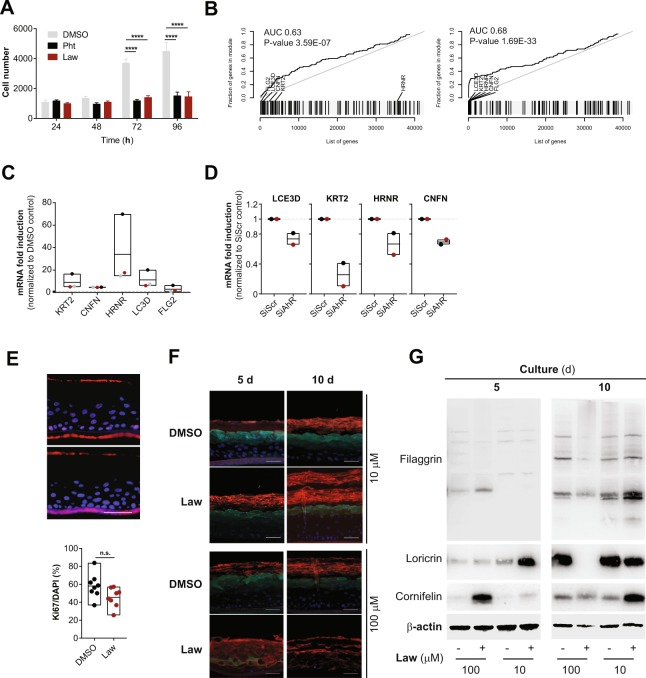
Lawsone stimulation modulates keratinocyte proliferation and differentiation. (A) Nuc red Live 647 positive HEK cells at different time points after stimulation with Lawsone (Law, 10 µM) and Phthiocol (Pht, 50 µM), compared to DMSO. (B) Epidermal differentiation complex and keratin gene enrichment of HEK cells after Lawsone stimulation (10 μM) and relative to TLR2 stimulation (Pam2CSK4, 0.236 μM) at (left) 4 h and (right) 24 h. Area under the curve (AUC), q-value and highly enriched genes are indicated. (C) KRT2, CNFN, HRNR, LCE3D and FLG2 expression of HEK cells after 24 h stimulation with Lawsone (10 µM) normalized to DMSO. Each color depicts results of the same individual. (D) LCE3D, KRT2, HRNRand CNFN expression on HEK cells transfected with AhR-siRNA (siAhR) or Scramble control (siScr) and further stimulated for 24 h with Lawsone (10 μM). Values are relative to siScr. Each color depicts results of the same individual. (E, top) Epidermal skin equivalents were stimulated for 5d with Lawsone (10 µM) or DMSO and stained with DAPI (blue) and the proliferation marker KI67 (purple). (E, bottom) Percentage of KI67 positive cells normalized to the total number of cells (DAPI). (F) Representative of an in vitro epidermis model experiment stained for Cornifelin (red) and Loricrin (green) and (G) protein expression of Filaggrin, Cornifelin and Loricrin at day 5 or 10 of culture after stimulation with 10 or 100 µM of Lawsone (blots were cropped from the same gel. Full unedited gels are provided in Supplementary Data). (A,C) Data from 3 independent experiments are shown. (D) Data from 2 independent donors. (E top, F,G) One representative experiment out of 2 is shown. (E) Pooled data from 2 different experiments is shown. (A) Mean + S.E.M., (C–E bottom) Floating bars, Mean Min to Max. (A) Two-way ANOVA with Fisher’s test, (C) One-way ANOVA with Dunn’s test. (E, bottom) Student’s t-test. *P < 0.05; **P < 0.01, ***P < 0.001, ****P < 0.0001.
Acknowledgments
This image is the copyrighted work of the attributed author or publisher, and
ZFIN has permission only to display this image to its users.
Additional permissions should be obtained from the applicable author or publisher of the image.
Full text @ Sci. Rep.