
IMAGE
Fig. S3
- ID
- ZDB-IMAGE-190815-18
- Publication
- Kang et al., 2019 - Ankrd45 Is a Novel Ankyrin Repeat Protein Required for Cell Proliferation
- All Figures
- Figures for Kang et al., 2019
Image
|
Figure Caption
Fig. S3
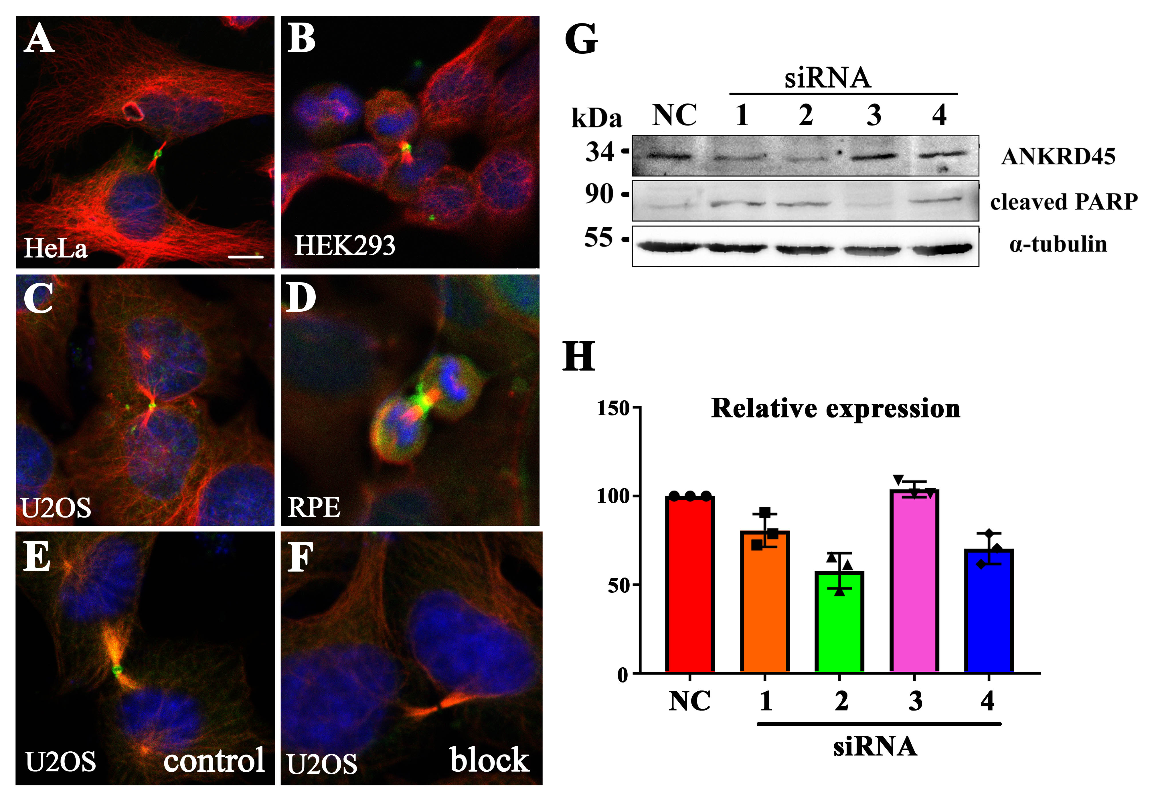
Localization of ANKRD45 in different cell lines
(A-D) Confocal images showing the localization of ANKRD45 in the midbody ring during cytokinesis of different cells as indicated. (E-F) Pre-incubation with synthesized ANKRD45 protein blocked the staining of ANKRD45 antibody in the midbody ring of U2OS cells. (G) Western blotting results showing the expression of ANKRD45 and cleaved PARP in control or four ANKRD45 siRNAs treated HeLa Cells. (H) Bar graph showing the relative expression level of ANKRD45 in control or siRNA treated HeLa cells. Scale bar: 5 μm.
Acknowledgments
This image is the copyrighted work of the attributed author or publisher, and
ZFIN has permission only to display this image to its users.
Additional permissions should be obtained from the applicable author or publisher of the image.
Full text @ Genes (Basel)