
Fig. S6
- ID
- ZDB-IMAGE-190814-27
- Publication
- Reuter et al., 2019 - Fgf3 is crucial for the generation of monoaminergic cerebrospinal fluid contacting cells in zebrafish
- All Figures
- Figures for Reuter et al., 2019
|
Fig. S6
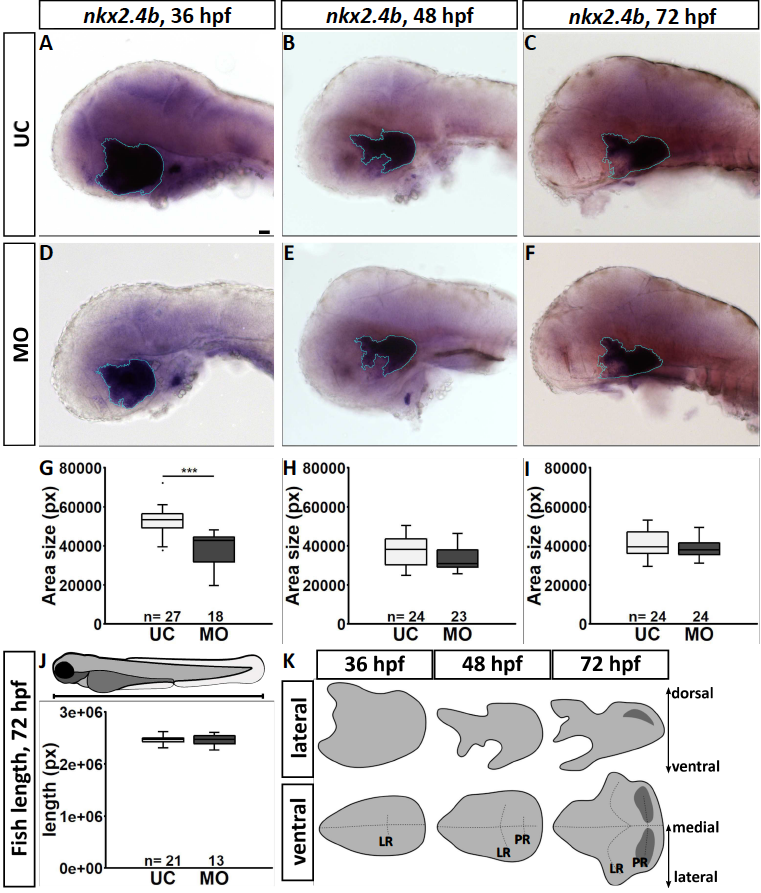
The shape of the hypothalamic nkx2.4b domain is altered in fgf3 morphants. (A-F) Light microscopic pictures showing expression of nkx2.4b in fgf3 morphants (MO) and uninjected control siblings (UC) at 36, 48 and 72 hpf visualised by RNA in situ hybridisation. Outlines of semi-automated measurement of hypothalamic area are highlighted in blue. Lateral views, anterior to the left. Scale bar = 30 μm. (G-I) Area measurements (pixels) in fgf3 morphants and control siblings of the nkx2.4b domain at 36, 48 and 72 hpf. (J) Total length measurements of fgf3 morphants and control siblings at 72 hpf. Tukey boxplots showing median, 25-75% percentile, IQR whiskers and outliers. n= number of analysed individuals. (K) Scheme illustrating the lateral and ventral silhouette of the nkx2.4b expressing hypothalamic domain at 36, 48 and 72 hpf. Dark grey indicates location of monoaminergic populations around the posterior recess (PR). Dashed lines show hypothalamic ventricular system with the (LR) lateral recess and the PR.