
|
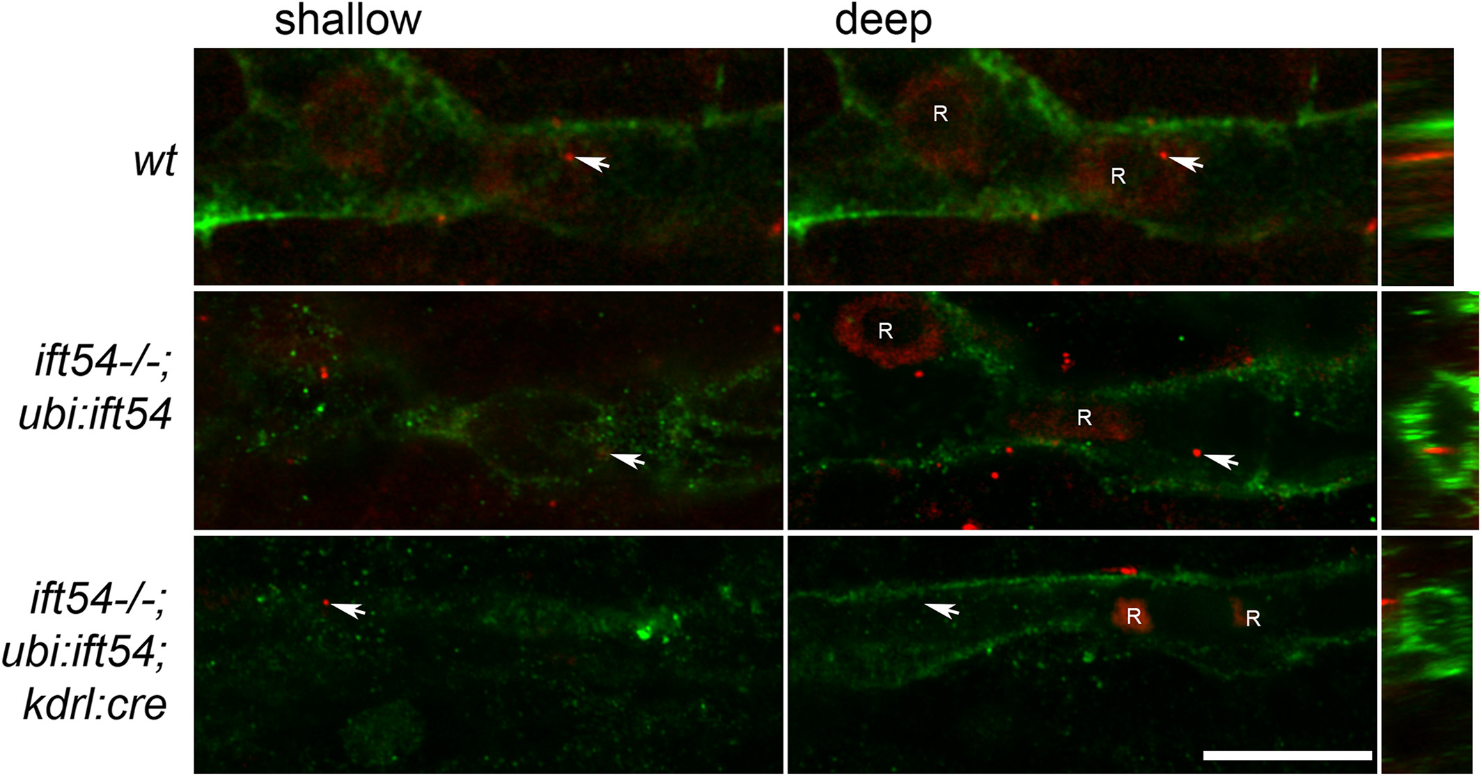
Fig. 7
Endothelial cilia are present in the juvenile caudal fin vascular plexus. Immunofluorescence confocal slices of anti‐acetylated tubulin (red)‐ and anti‐GFP (green)‐stained caudal fin vascular plexus of 11‐dpf Tg(fli1a:LIFEACT‐clover)sh467 /+ (wt) (n = 15), 13‐dpf ift54 tp49; Tg(ubi:loxP‐ift54‐loxP‐myr‐mcherry,myl7:EGFP)sh488 /+;Tg(fli1a:LIFEACT‐clover)sh467 /+ (ift54−/−; ubi:ift54) (n = 4), and 13‐dpf ift54 tp49; Tg(ubi:loxP‐ift54‐loxP‐myr‐mcherry,myl7:EGFP)sh488/+;Tg(kdrl:cre)s898 /+;Tg(fli1a:LIFEACT‐clover)sh467 /+ (ift54−/−;ubi:ift54;kdrl:cre) (n = 4). Slices at shallow and deep levels are shown along with orthogonal views resliced through the confocal stack at the level of cilia marked by arrows. Autofluorescent red blood cells (R) are visible in the vessel lumen. GFP, green fluorescent protein; WT, wild‐type. Scale bar = 10 μm