
|
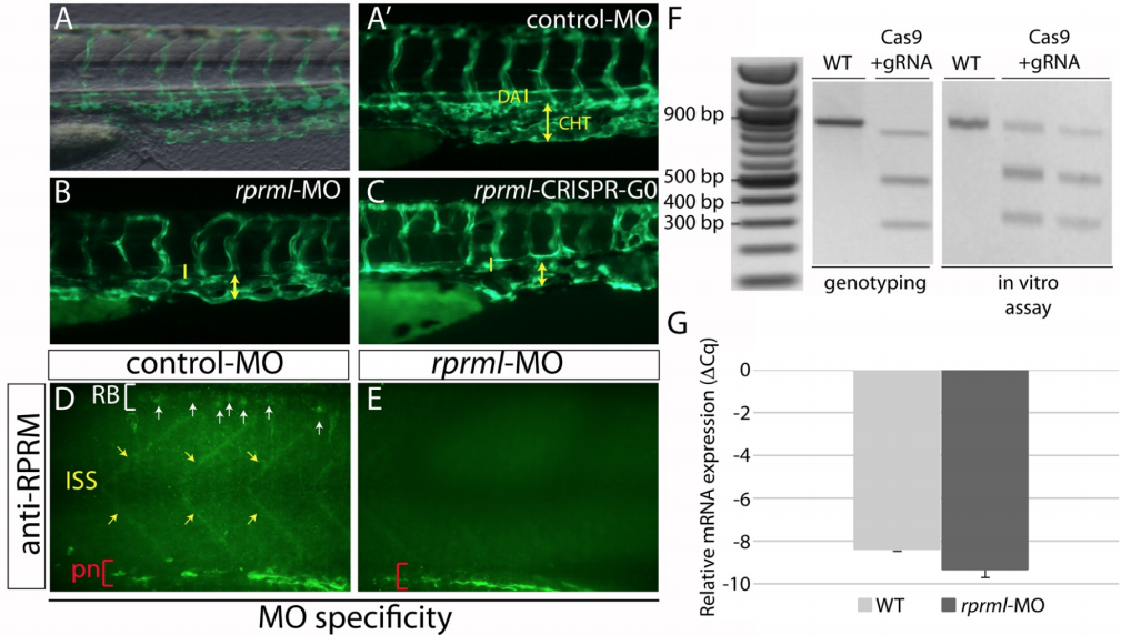
Fig. S2
Similar phenotypes are obtained by disruption of rprml, either by CRISPR-Cas9 mutation or injection of antisense MOs. (A-E) lateral views of (A-C) live transgenic Tg(fli1:GFP) or (D-E) anti-RPRM IHC stained embryos at 48 hpf visualized by fluorescence and confocal microscopy, respectively. (A-A’) Normal vascular morphology is observed in control-MO-injected embryos. (B-C) Embryos injected with MOs or CRISPR-Cas9 targeting rprml exhibit reduced caudal hematopoietic tissue (CHT) territory (yellow double arrows). The yellow line represents the dorsal aorta (DA). (D-E) Rprm/Rprml protein expression is effectively blocked by rprml-MO. A human anti-RPRM/RPRML antibody labeled the Rohon-Beard neurons (RB, white arrows), the inter-somitic spaces (ISS, yellow arrows) and the pronephric tubule (pn, red brackets). A drastic reduction in immunoreactivity is observed in rprml MO-injected embryos compared to control MO-injected embryos. (F) Left panel: rprml CRISPR-Cas9 embryo genotyped by T7 endonuclease assay shows INDEL mutations of the expected sizes (500bp, 350bp). Right panel: in vitro DNA cleavage activity assay showing cropped areas containing Cas9-cleaved DNA bands of the expected sizes for rprml. A 1 kb DNA ladder was used as a molecular weight marker. (G) qPCR showing p53 relative expression in wild type and rprml MO specimens. Data shown as ΔCq ± SEM, (CI 95% = -8.41 ± 0.135 for wild type controls; CI 95% = -9.36 ± 0,65 for rprml morphants).