
|
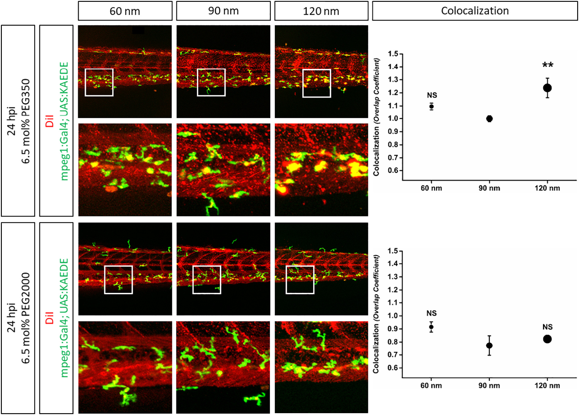
Fig. 5
Influence of liposome size and different PEG modification on macrophage clearance in zebrafish. PEG350 or PEG2000 modified and DiIlabeled liposomes with three different sizes (60 nm, 90 nm, and 120 nm) were injected into the blood circulation of zebrafish 2 days post fertilization. Confocal images were acquired 24 h post injection (hpi). Colocalization of green fluorescent macrophages (KAEDE) and DiI labeled liposomes was quantitated using normalized overlap colocalization coefficients. Values are means ± SEM, n = 5. not significant (NS) or **P < 0.05 (ANOVA and Bonferroni correction) as compared to 90 nm sized liposomes.
Reprinted from Nanomedicine : nanotechnology, biology, and medicine, 17, Sieber, S., Grossen, P., Uhl, P., Detampel, P., Mier, W., Witzigmann, D., Huwyler, J., Zebrafish as a predictive screening model to assess macrophage clearance of liposomes in vivo, 82-93, Copyright (2019) with permission from Elsevier. Full text @ Nanomedicine