
|
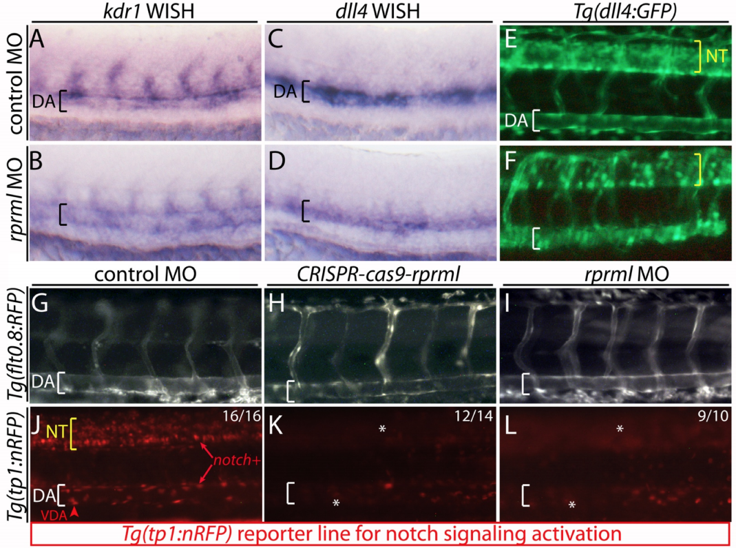
Fig. S6
lack of rprml hinders normal activation of the Notch signaling pathway. (AD) Lateral views of 24 hpf analyzed by WISH for (A-B) arterial endothelial cells (kdr1) and (C-D) vascular arterial progenitor cells (dll4). (E-L) Fluorescent microscopy images of transgenics: (E-F) Tg(dll4:GFP), (G-I) Tg(flt0.8:RFP) and (J-L) Tg(Tp1:nRFP). Brackets indicated the positioning of the dorsal aorta (DA). (K-L) Asterisks indicated reduced Notch activity in CRISPR-Cas9-rprml and/or rprml-MO injected embryos (DA). The number of embryos with the phenotype shown as a fraction of the total number of embryos examined is indicated in the top right corner in J-L.