
Image
|
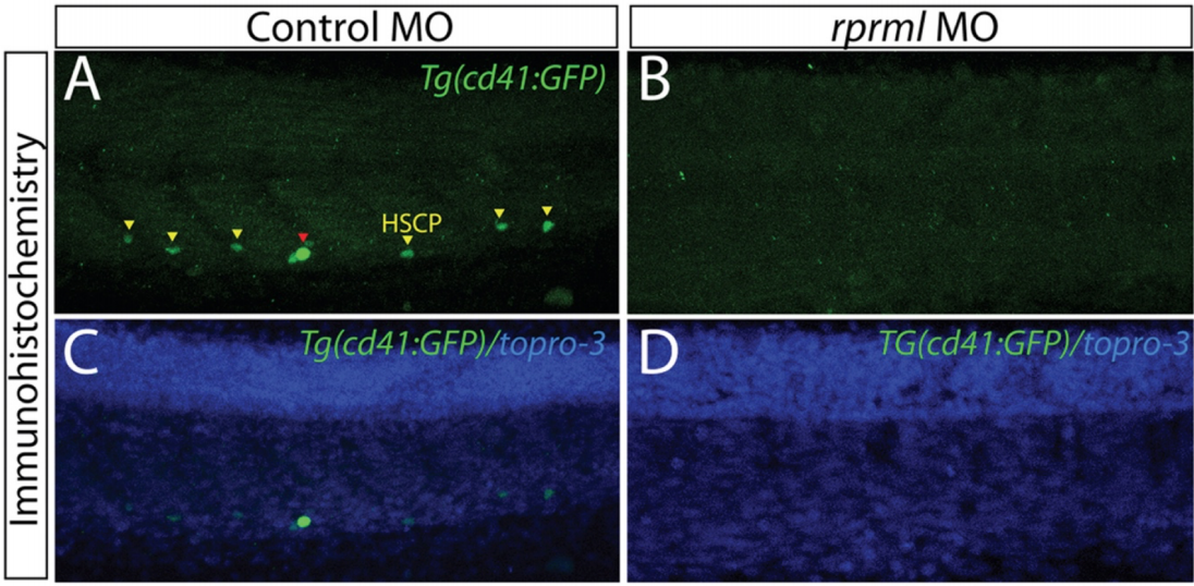
Figure Caption
Fig. S4
CD41 expression is reduced in the CHT of rprml morphants. (A-D) Lateral view of the trunk with anterior to the left. (A-B) Whole mount immunohistochemistry/immunofluorescence showing Tg(CD41:GFP) expression pattern at 54 hpf. Staining with anti-GFP indicates expression of HSPC (yellow arrows) in the CHT (green bracket). (C-D) topro-3 staining (blue) shows the localization of the nuclei.
Acknowledgments
This image is the copyrighted work of the attributed author or publisher, and
ZFIN has permission only to display this image to its users.
Additional permissions should be obtained from the applicable author or publisher of the image.
Full text @ Sci. Rep.