
|
Fig. 1
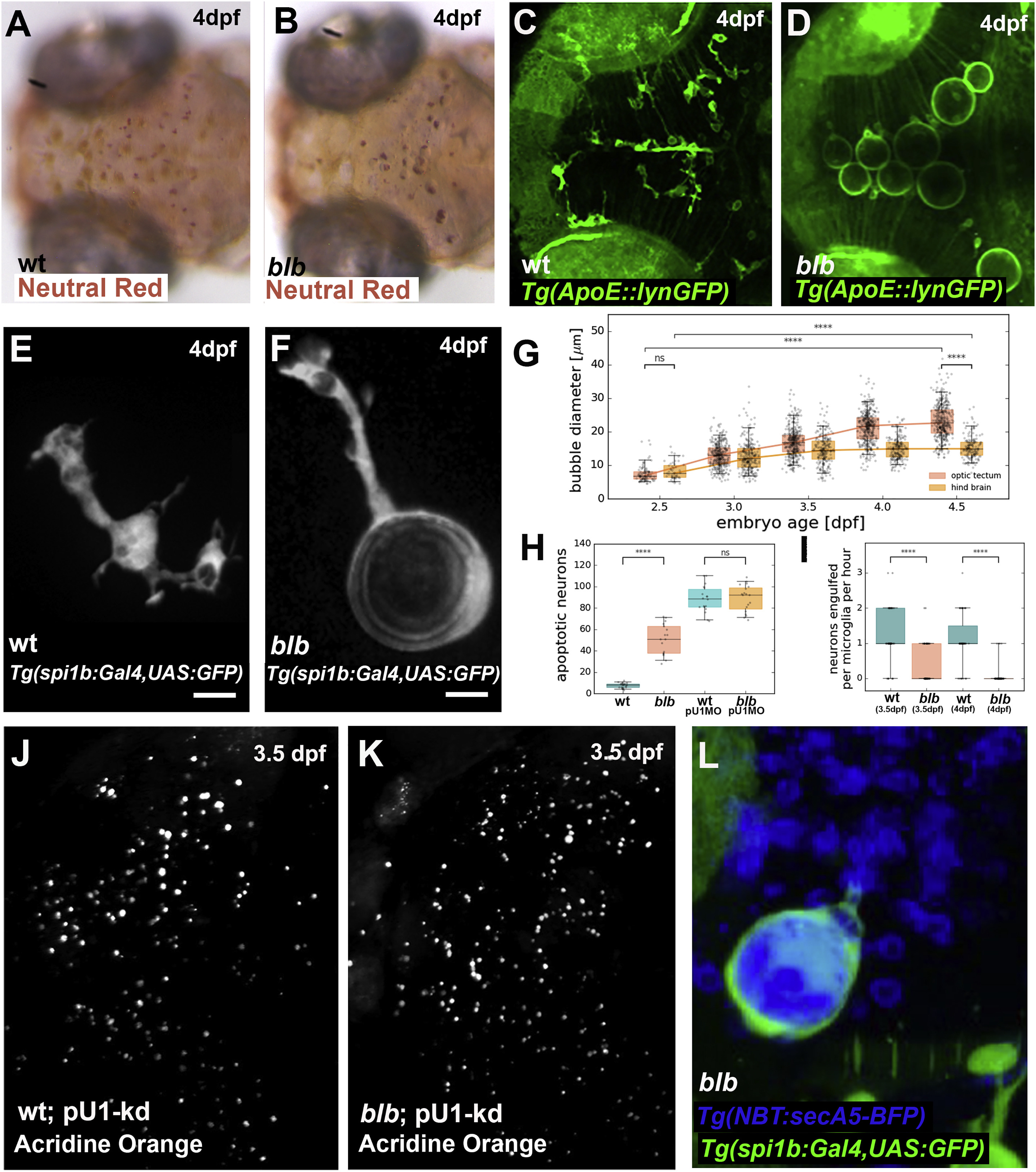
Characterization of bubblebrain, a Mutant with Bloated Microglia
(A and B) Neutral red staining in a 4 dpf wild-type (A) and blbNY007 (B) embryos.
(C and D) Microglia labeled with Tg(Apo-E:lynGFP) in a 4 dpf wild-type (C) and blbNY007 (D) embryo.
(E and F) Microglia cell labeled with Tg(spi1b:Gal4,UAS:GFP) in a 4 dpf wild-type (E) and blbNY007 (F) embryo. Scale bar, 10 μm.
(G) Quantification of the “bubble” size in blbNY007 embryos from 2.5 to 4.5 dpf in optic tectum (OT, red plots) and in the hindbrain (HB, orange plots). Whiskers: 5th/95th percentile, p value < 0.0001.
(H) Quantification of apoptotic neurons visualized by Acridine Orange in wild-type (n = 20), blbNY007 (n = 17) and in wild-type (n = 18) and blbNY007 (n= 19) injected with pU1 morpholino to deplete microglia in 3.5 dpf embryos; representative images in (J) and (K). Whiskers: 5th/95th percentile, p value < 0.0001.
(I) Quantifications of phagocytic events per cell per hour in wild-type (n = 50; n = 47) and blbNY007 (n = 61; n = 77) embryos at 3.5 dpf and 4 dpf, respectively. Whiskers: 5th/95th percentile, p value < 0.0001.
(J and K) Dorsal view of a 3.5 dpf wild-type (J) and blbNY007 (K) embryo injected with pU1 morpholino to deplete microglia and stained with Acridine orange to visualize apoptotic neurons; quantifications in (H).
(L) Microglia cell labeled with Tg(mpeg:GFP-caax) and Tg(NBT:secA5-BFP) to visualize apoptotic neurons in a 4 dpf blbNY007 embryo.
Reprinted from Developmental Cell, 49(1), Villani, A., Benjaminsen, J., Moritz, C., Henke, K., Hartmann, J., Norlin, N., Richter, K., Schieber, N.L., Franke, T., Schwab, Y., Peri, F., Clearance by Microglia Depends on Packaging of Phagosomes into a Unique Cellular Compartment, 77-88.e7, Copyright (2019) with permission from Elsevier. Full text @ Dev. Cell