IMAGE

Figure 3

ID
ZDB-IMAGE-190723-409
Source
Figures for Stanic et al., 2019
Image
Figure Caption

Figure 3

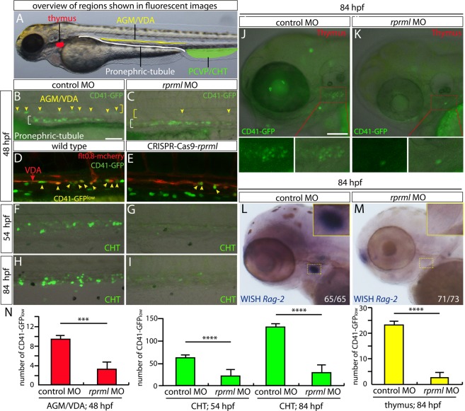
rprml is required for specification of hematopoietic precursor/stem cells (HSPCs) from the hemogenic endothelium at the ventral dorsal aorta. (A) Lateral view of zebrafish embryo with anterior to the left, showing overview of regions magnified in the fluorescent imaging (BK) and WISH (L,M) panels. Red region denotes the left thymic lobe, yellow region the anterior gonad mesonephros/ventral dorsal aorta (AGM/VDA), green region the posterior caudal vein plexus/caudal hematopoietic tissue (PCVP/CHT) and white region the pronephric-tubule. (BM) Lateral views of zebrafish embryos with anterior to the left at denoted developmental stages (48, 54 and 84 hpf). Loss-of function of rprml impairs normal formation of CD41-GFPlow+ HSPCs (yellow arrows in BE) originating from the AGM/VDA (BE, red bar graphs in N) and their migration to the CHT (FI, green bar graphs in N) and thymus (J,K, yellow bar graphs in N). (D,E) Double transgenic Tg(flt0.8:mcherry;CD41:GFP) CRISPR-Cas9-rprml injected embryos show the same decrease in HSPCs originating from the AGM/VDA as rprml-morphant embryos. (L,M) WISH showing that rprml-morphant embryos show a remarkable absence of rag2 (lymphocyte marker) in the thymus. Statistical significance was determined using two-tailed unpaired Student’s t-test. ***P ≤ 0.001, ****P ≤ 0.0001. Scale bars, 50 μm (BI) and 100 μm (JM).

Acknowledgments
This image is the copyrighted work of the attributed author or publisher, and ZFIN has permission only to display this image to its users. Additional permissions should be obtained from the applicable author or publisher of the image. Full text @ Sci. Rep.