IMAGE

Figure 1

ID
ZDB-IMAGE-190723-405
Source
Figures for Stanic et al., 2019
Image
Figure Caption

Figure 1

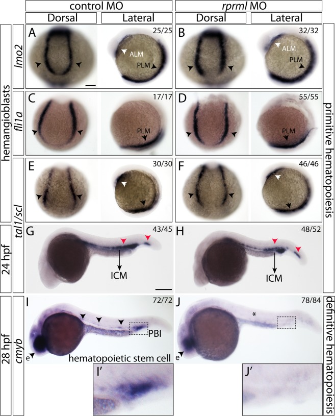
rprml is dispensable for primitive hematopoiesis but required for HSPC formation. (AF) Dorsal and lateral views of 10 somite-stage zebrafish embryos analyzed by WISH. White and black arrowheads represent the anterior- (ALM) and posterior-lateral plate mesoderm (PLM), respectively. (A,C,E,G,I) control MO-injected embryos; (B,D,F,H,J) rprml MO-injected embryos. (AF) Expression patterns of typical hemangioblast markers: (A,B) lmo2, (C,D) fli1a, (E,F) tal1/scl1. (G,H) Lateral view of 24 hpf control MO and rprml MO-injected embryos stained by WISH for tal1/scl1. Black arrows indicate the intermediate cell mass (ICM) and red arrowheads normal expression pattern for tal1/scl1. (I,J) 28 hpf embryos stained by WISH for cmyb comparing controls versus rprml MO-injected embryos. Black arrowheads indicate normal expression pattern for cmyb. rprml morphant embryos retain retinal expression of cmyb (eye, e). The number of embryos with the phenotype shown as a fraction of the total number of embryos examined is indicated in the top right corner in (AJ). Scale bars, 100 μm (AF) and 200 μm (GJ).

Acknowledgments
This image is the copyrighted work of the attributed author or publisher, and ZFIN has permission only to display this image to its users. Additional permissions should be obtained from the applicable author or publisher of the image. Full text @ Sci. Rep.