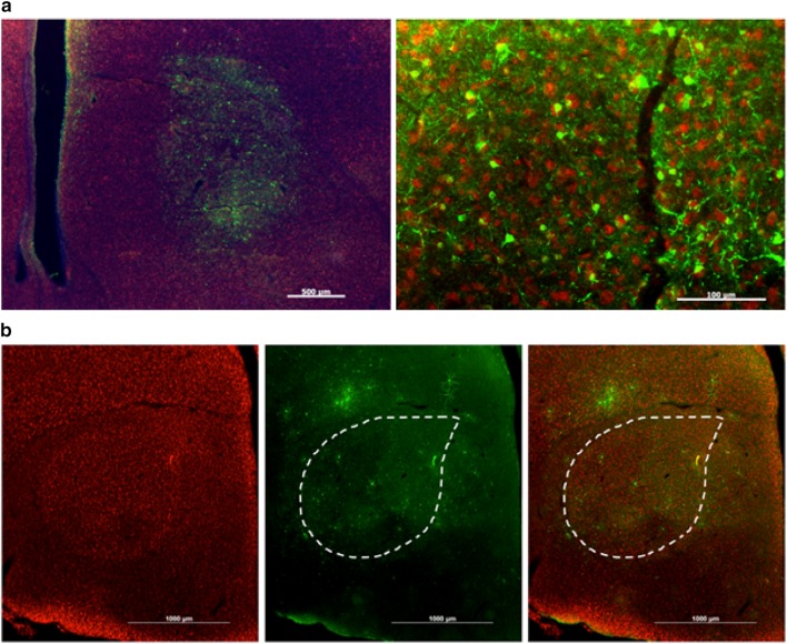
Virovek AAV2/5 is effective in transducing Area X neurons, whereas AAV2/9 is not. a Low magnification photomicrograph (left) shows that AAV2/5 can transfect a delimited region of the brain. Some transfection is seen dorsal of the striato-nidopallial border, likely the result of mis-targeting of the injection. Scale bar 500 μm. Higher magnification, right, reveals a high transfection rate. Scale bar 100 μm. b Anti-NeuN antibody signals (red) reveal Area X in left-most panel that is outlined in the middle and right panels. AAV2/9 injection in Area X leads to GFP-positive signals (green, middle panel) in cells that are diffusely scattered around the injection site. Images are merged in the right panel
Acknowledgments
This image is the copyrighted work of the attributed author or publisher, and
ZFIN has permission only to display this image to its users.
Additional permissions should be obtained from the applicable author or publisher of the image.
Full text @ J. Comp. Physiol. A
Your Input Welcome
Thank you for submitting comments. Your input has been emailed to ZFIN curators who may contact you if
additional information is required.
Oops. Something went wrong. Please try again later.