IMAGE

Figure 1

ID
ZDB-IMAGE-190723-2263
Source
Figures for Dütting et al., 2011
Image
Figure Caption

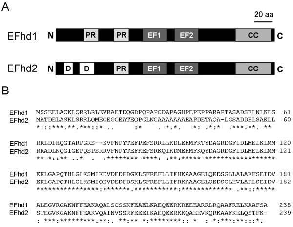
Figure 1

Predicted secondary structures and sequence comparisons of EFhd2 and EFhd1. a) Predicted secondary structures of murine EFhd1 and EFhd2 (240 aa) (http://elm.eu.org). PR: proline-rich elements (potential SH3 domain-binding sites), EF: EF-hands, CC: coiled-coil domain, D: disordered region.

b) Aligment of murine EFhd1 (Q9D4J1) and EFhd2 (Q9D8Y0) sequences using ClustalW2 (http://www.ebi.ac.uk/clustalw). Amino acid (aa) positions are marked on the right. "*", identical aa, ":", conserved aa, "." semi conserved aa.

Acknowledgments
This image is the copyrighted work of the attributed author or publisher, and ZFIN has permission only to display this image to its users. Additional permissions should be obtained from the applicable author or publisher of the image. Full text @ Cell Commun. Signal.