IMAGE

Fig 1

ID
ZDB-IMAGE-190723-1186
Source
Figures for Murdoch et al., 2019
Image
Figure Caption

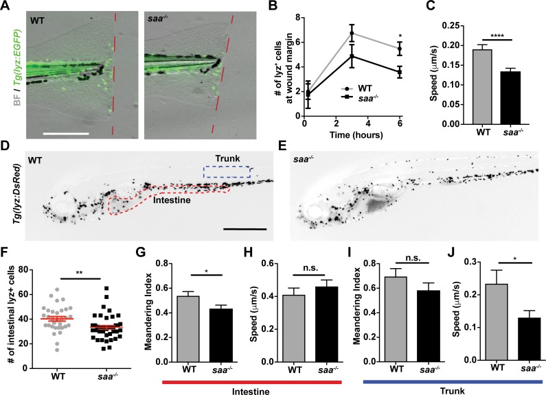
Fig 1 Saa mediates neutrophil behavior <italic>in vivo</italic>.

(A-B) Imaging and quantification of lyz:EGFP+ neutrophils recruited to tail wound margin over 6 hours following caudal fin amputation (dashed red line indicates wound margin; scale bar = 250 μm) (n ≥ 24 larvae / genotype at 6 hour time point). (C) Measurement of lyz:DsRed+ neutrophil speed from time-lapse imaging in caudal fin tissue over 6 hour period following amputation (n = 4 larvae / genotype, 87–112 cells tracked / genotype). (D-E) Representative images of 6 dpf Tg(lyz:DsRed) WT and saa-/- larvae (scale bar = 500 μm). (F) Enumeration of intestine-associated lyz:EGFP+ cells in 6 dpf larvae (n = 32–40 larvae / genotype). (G-J) Quantitative analysis of lyz:EGFP+ neutrophil behavior from time-lapse imaging of distinct anatomical compartments (intestine and trunk, ROIs in panel D) in 6 dpf larval zebrafish (6 larvae / genotype, ≥ 23 cells analyzed / genotype / tissue). Data analyzed by t-test. For panel B, statistical comparisons were performed within each time point. Data are presented as mean ± SEM. * p < 0.05, ** p < 0.01, *** p < 0.001, **** p < 0.0001.

Figure Data
Acknowledgments
This image is the copyrighted work of the attributed author or publisher, and ZFIN has permission only to display this image to its users. Additional permissions should be obtained from the applicable author or publisher of the image. Full text @ PLoS Pathog.