
Fig. 5
- ID
- ZDB-IMAGE-190718-41
- Genes
- Publication
- Marra et al., 2019 - Prostaglandin signaling regulates renal multiciliated cell specification and maturation
- All Figures
- Figures for Marra et al., 2019
|
Fig. 5
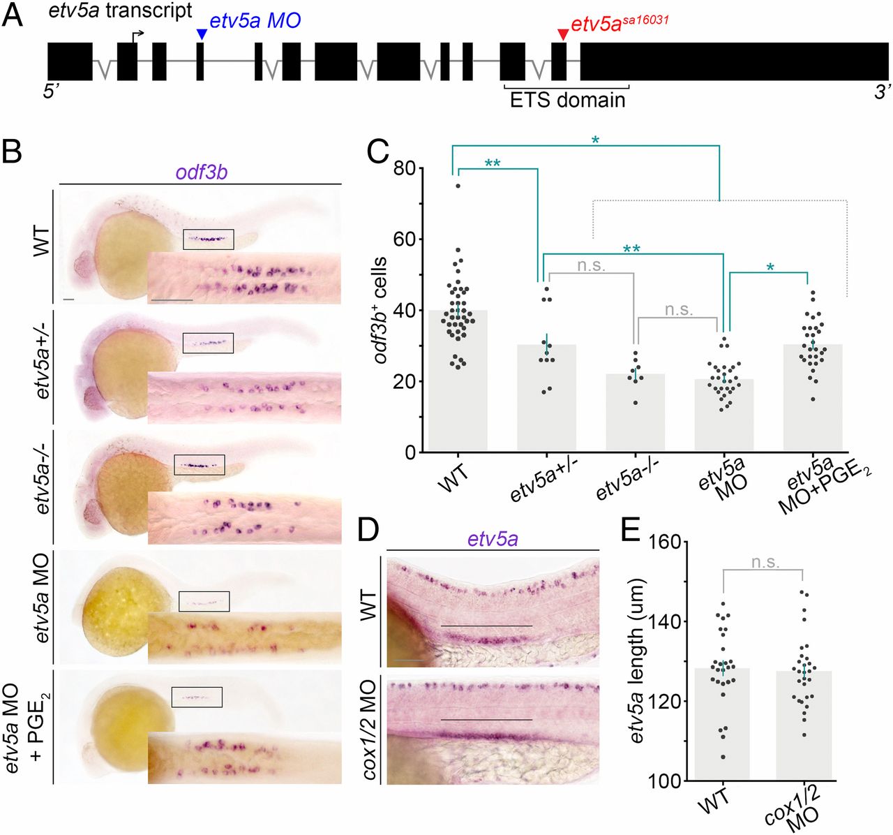
Prostaglandin signaling acts downstream of etv5a during MCC genesis. (A) Schematic of the etv5a locus with locations of the splice MO (blue arrowhead) and the etv5asa16031mutation (red arrowhead). The forward arrow in exon 2 depicts the ATG start site. (B) WISH at the 28 ss on representative genotype-confirmed WT embryos, etv5a morphants, and etv5a morphants treated with dmPGE2. Embryos are imaged in a lateral view, and the Insets correspond to a dorsal view of the pronephros. (Scale bar, 50 μm.) (C) Quantification of odf3b+ cells at the 28 ss. Each dot represents the total number of maturing renal MCCs in one embryo. Data are represented as mean ± SEM; significance was determined by ANOVA. (D) Lateral view of the etv5a domain (black line) in the pronephros of representative WT and cox1/2 morphants. (Scale bar, 50 μm.) (E) Quantification of absolute etv5a length in WT versus cox1/2 morphants. Each dot represents the etv5a domain length in one nephron. Data are represented as mean ± SEM; significance was determined by Student’s t test, where *P < 0.0001, **P < 0.006, n.s., not significant.