
Image
|
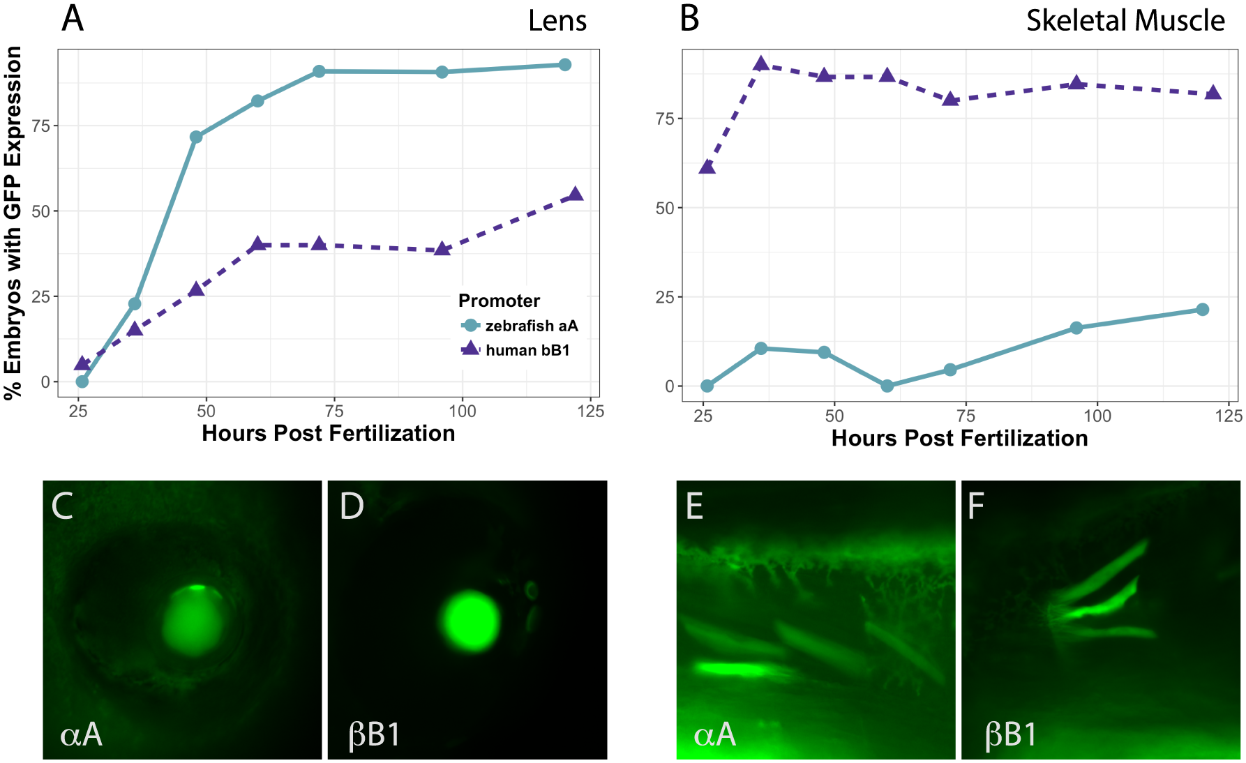
Figure Caption
Fig. 7
Timecourse of promoter activity in lens (A) and skeletal muscle (B) in all combined embryos.
Zebrafish αA-crystallin promoter (circles) produced lens expression in a larger proportion of embryos by 36 hpf and drove GFP expression in over 85% of embryos by 72 hpf. The human βB1-crystallin promoter (triangles) drove surprisingly abundant expression in zebrafish skeletal muscle, but was less active in lens. Between 11 and 66 embryos were observed for each timepoint. Images C-F show representative examples of GFP expression with each promoter as indicated in lens (C and D) and skeletal muscle (E and F).
Acknowledgments
This image is the copyrighted work of the attributed author or publisher, and
ZFIN has permission only to display this image to its users.
Additional permissions should be obtained from the applicable author or publisher of the image.
Full text @ PLoS One