
Fig. 2
- ID
- ZDB-IMAGE-190626-10
- Genes
- Publication
- Gerber et al., 2019 - The HMG box transcription factors Sox1a and b specify a new class of glycinergic interneurons in the spinal cord of zebrafish embryos
- All Figures
- Figures for Gerber et al., 2019
|
Fig. 2
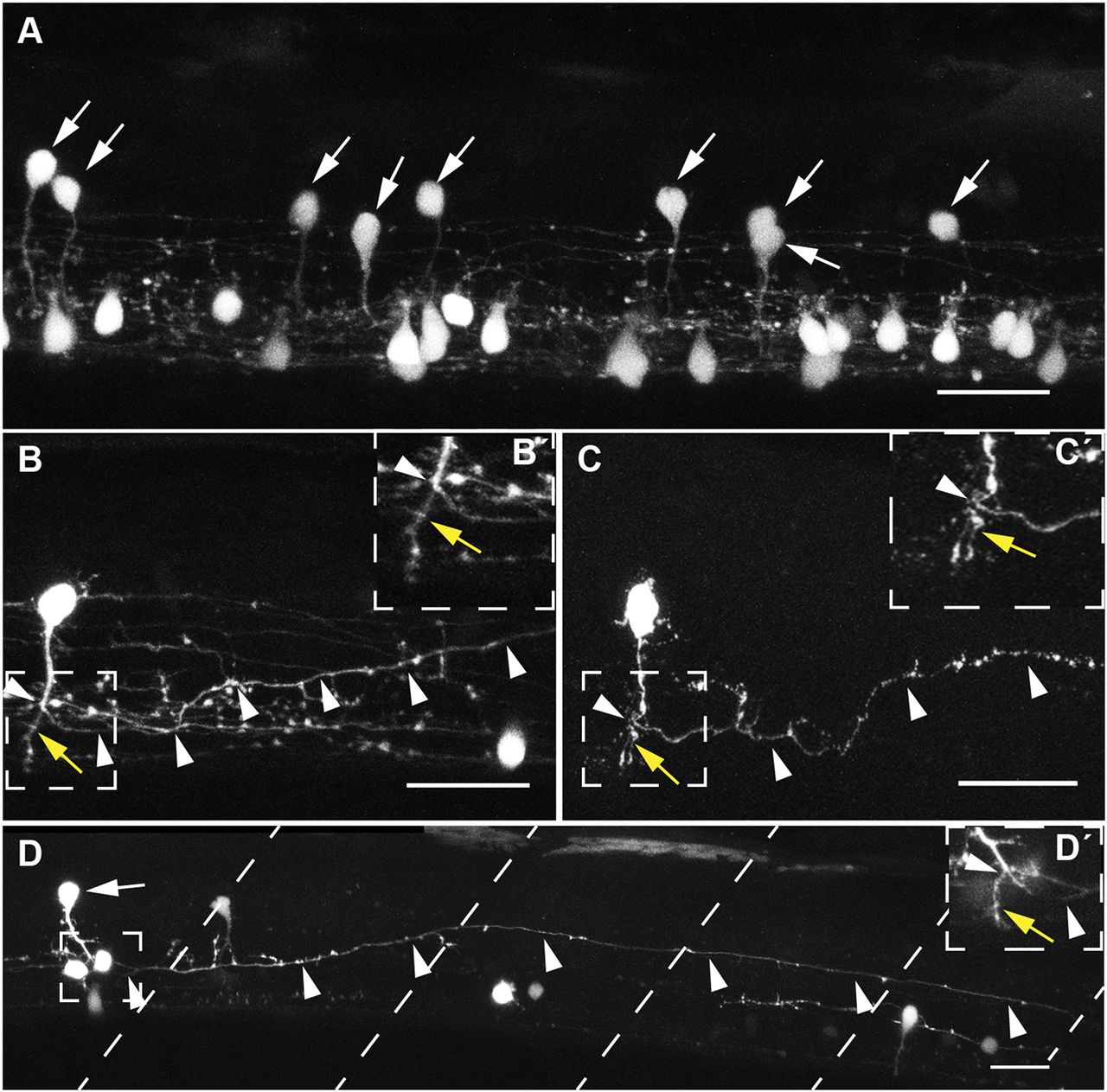
Morphology of V2s neurons. (A) Spinal cord of a sox1a:eGFP embryo with eGFP+ neurons in the V2 domain (arrows) at 60 hpf over a section of three somites above the yolk extension. Ventrally located sox1a:eGFP+ cells without arrows are KA′ and KA″ neurons. sox1a:eGFP+ V2 neurons show an oval-shaped soma at an intermediate spinal cord position and an axon extending ventrally towards the floor plate. (B,B′) sox1a:eGFP+ V2 neuron in a mosaic eGFP knockout embryo at 60 hpf. The ventrally extending axon branches over the floorplate into a long axon descending ipsilaterally (arrowheads) and a short axon branch ending ventrally (B′, yellow arrow). (C,C′) A sox1a+ neuron labelled transiently by TgBAC(sox1a:eGFP) at 48 hpf with an oval-shaped soma and ventrally extending axon that branches into a short axon ending ventrally (C′, yellow arrow) and a long axon descending and rising to an intermediate DV level of the spinal cord (arrowheads). (D,D′) The sox1a:eGFP+ V2 neuron (arrow) extends a long axon ipsilaterally over five somites (somite boundaries are indicated by dashed lines). (D′) Different focal plane showing the short axon branch (yellow arrow) ending ventrally and the main axon branching and descending (arrowheads). Dorsal is upwards; anterior is leftwards. Data are derived from at least two independent experiments. Scale bars: 25 µm in A-C; 100 µm in D.