Fig. 1
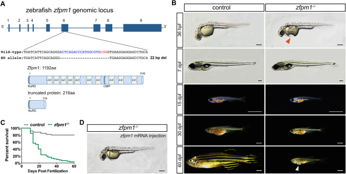
Generation of Zfpm1 knock out zebrafish line. (A): Upper panel, the zebrafish Zfpm1 genomic locus and Cas9/sgRNA targeting site. Deletions in ∆22 allele are shown as dashes. Lower panel, schematic representations of the domain structure of the wild-type zebrafish Zfpm1 protein and truncated protein derived from the ∆22 allele. (B) Live images of control and Zfpm1-/- zebrafish at designated time points. Lateral view, anterior to the left. Red arrowhead indicates the depression on the yolk of Zfpm1-/-embryo, white arrowhead indicates the bulge on the ventral side of Zfpm1-/-fish. For 36hpf embryos and 7dpf larvae, scale bar: 200 µm. For animal from 15 dpf to 60 dpf, scale bar: 2 mm. (C) Representative Kaplan-Meier plot for Zfpm1-/- fish and clutchmates from one of three independent experiments. 80 total Zfpm1-/- animals and 88 total siblings were followed. P < 0.0001, Mantel-Cox test. (D) Injection of Zfpm1 mRNA rescue the morphological defects in Zfpm1-/- embryos. Scale bar: 200 µm. (For interpretation of the references to color in this figure legend, the reader is referred to the web version of this article.)
Reprinted from Developmental Biology, 446(2), Yang, Y., Li, B., Zhang, X., Zhao, Q., Lou, X., The zinc finger protein Zfpm1 modulates ventricular trabeculation through Neuregulin-ErbB signalling, 142-150, Copyright (2019) with permission from Elsevier. Full text @ Dev. Biol.

