
Fig. 6
- ID
- ZDB-IMAGE-190618-68
- Publication
- James et al., 2019 - Intestinal dysmotility in a zebrafish (Danio rerio) shank3a;shank3b mutant model of autism
- All Figures
- Figures for James et al., 2019
|
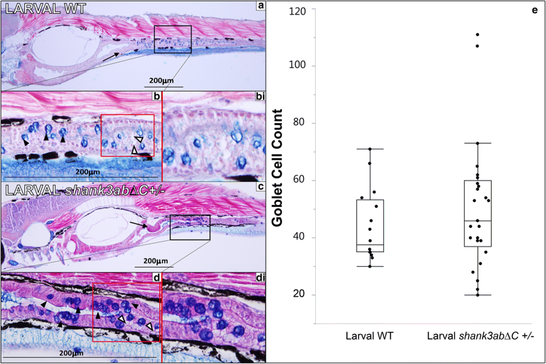
Fig. 6
Histology of larval WT and shank3abΔC+/− mutants (n = 25 for shank3abΔC +/−n = 15 for WT). Black boxes in a and c indicate regions shown at 40× magnification, while red boxes in b and d indicate regions of magnified insets in bi and di. a 10× magnification of longitudinal sections through 7 dpf WT fish (anterior left to posterior right); sections were stained with alcian blue and Eosin B. Sections show a well-defined polarized epithelium, with folding beginning in the intestinal bulb and extending into the upper intestine (black arrow). b The goblet cells are stained dark blue (black arrowheads) and intestines (stained purple) are clearly visible, while mucous production can be seen in the luminal space, stained light blue. bi This magnified inset from b shows enterocytes with large supranuclear vesicles (white arrowheads). c 10× magnification of 5-μm sections through 7 dpf shank3abΔC +/− mutants. Mucous production can be seen in luminal space shown in 40× magnification of d, along with goblet cells (black arrowheads) and intestinal lumen. di Similar to what is seen in WT fish, shank3abΔC+/− mutants display enterocytes with large supranuclear vesicles (white arrowheads). e Comparison of WT and shank3abΔC +/− mutant goblet cell count; no significant difference was found at 7 dpf