
Fig. 2
- ID
- ZDB-IMAGE-190618-64
- Antibodies
- Publication
- James et al., 2019 - Intestinal dysmotility in a zebrafish (Danio rerio) shank3a;shank3b mutant model of autism
- All Figures
- Figures for James et al., 2019
|
Fig. 2
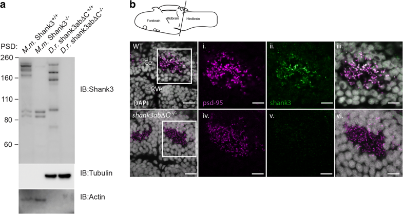
Zebrafish Shank3ab protein shares complex isoform expression with mammals and shows expression in the cerebellum. a Shank3 western blots of postsynaptic densities (PSD) isolated from mouse (M.m) and zebrafish (D.r.) show similar complex isoform expression. Molecular marker weight is expressed in kilodaltons. Tubulin and actin expression were used as loading controls for zebrafish and mouse immunoblots, respectively. Loss of Shank3ab staining in IHC and the western blot supports the immunoreactive specificity of Shank3 antibody. b Shank3ab protein is expressed as distinct overlapping puncta with PSD-95 in the cerebellum from larvae 6 days post-fertilization. Transverse cerebellar sections (see diagram) with enlarged insets show neuropil area dorsal to the rhombencephalic ventricle (RVe). Scale bar represents 10 μm for the first column and 5 μm for the insets (i–vi)