
Fig. 3
- ID
- ZDB-IMAGE-190618-58
- Genes
- Publication
- Li et al., 2018 - Znfl1s are essential for patterning the anterior-posterior axis of zebrafish posterior hindbrain by acting as direct target genes of retinoic acid
- All Figures
- Figures for Li et al., 2018
|
Fig. 3
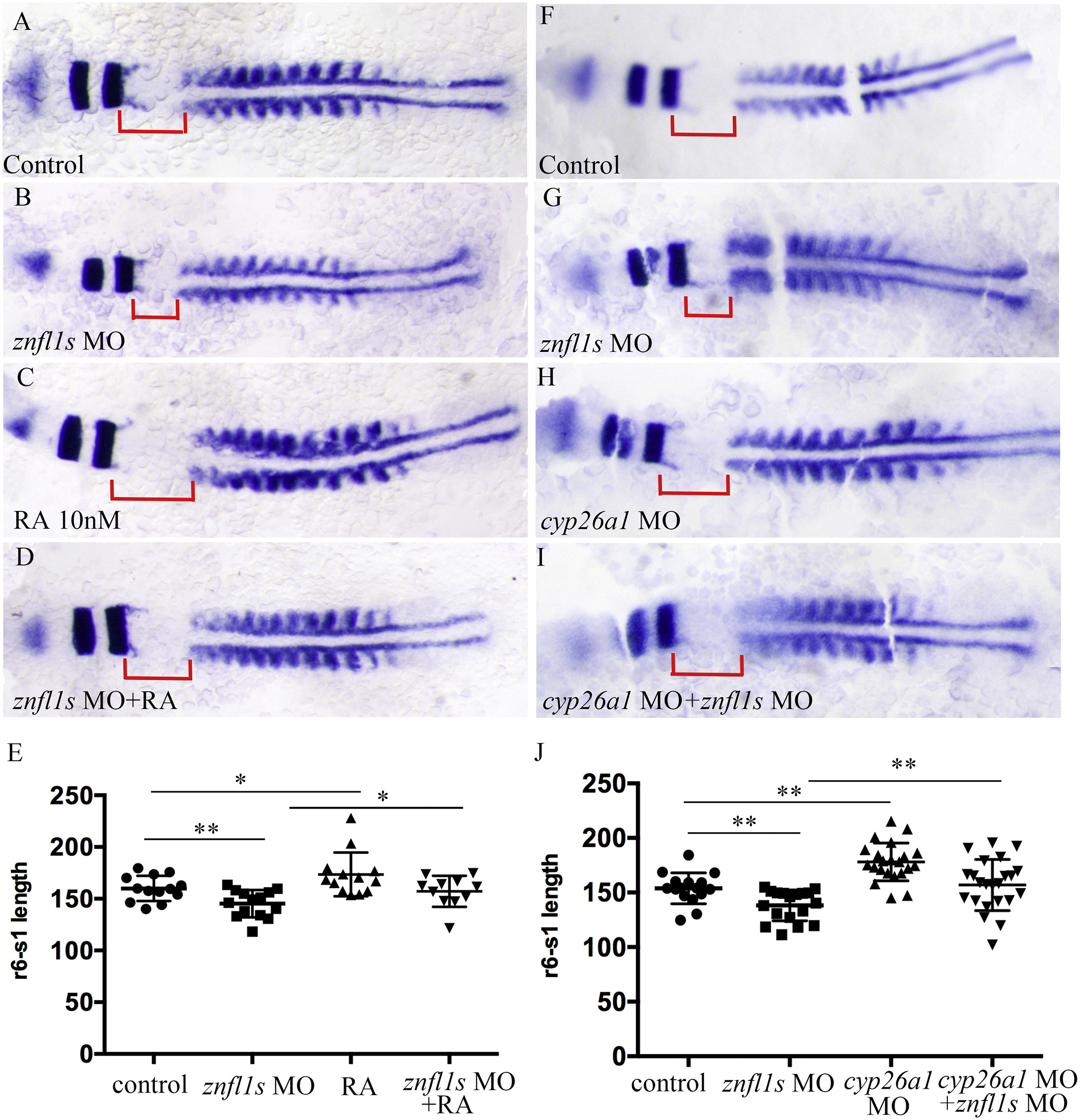
Increasing RA signaling rescues the defective posterior hindbrain of znfl1s morphants. Flat mount embryos at 11–12-somites stage are shown anterior left (A–D, F–I). At 11–12 somitestage, the length of r6-s1 were measured from the control embryos (A, F), znfl1s morphants (B, G), 10 nM RA treated embryos (C), znfl1s MO microinjected plus RA treated embryos (D), cyp26a1morphants (H), cyp26a1 MO plus znfl1s MO microinjected embryos (I). The data about the length of r6-s1 derived from A–D and F–I are shown in scatter diagrams E and J, respectively. The red line shows the length of r6-s1. **p < 0.01; * p < 0.05.
Reprinted from Mechanisms of Development, 155, Li, J., Zhao, Y., He, L., Huang, Y., Yang, X., Yu, L., Zhao, Q., Dong, X., Znfl1s are essential for patterning the anterior-posterior axis of zebrafish posterior hindbrain by acting as direct target genes of retinoic acid, 27-33, Copyright (2018) with permission from Elsevier. Full text @ Mech. Dev.