
IMAGE
Fig. S5
- ID
- ZDB-IMAGE-190530-17
- Publication
- Konjikusic et al., 2018 - Mutations in Kinesin family member 6 reveal specific role in ependymal cell ciliogenesis and human neurological development
- All Figures
- Figures for Konjikusic et al., 2018
Image
|
Figure Caption
Fig. S5
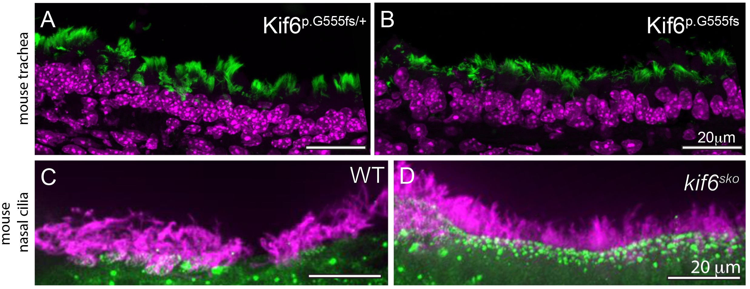
Immunofluorescence (IF) of Kif6 mutant multiciliated tissues in mouse and zebrafish.
(A-B) Immunofluorescence of trachea sections in Kif6p.G555fs/+ and Kif6p.G555fs mice showing no apparent cilia defects present in trachea of Kif6 mutant mice. Acetylated tubulin (green) marking cilia, DAPI-stained nuclei (magenta) (C-D) Representative IF of zebrafish nasal pit cilia shows typical cilia in kif6 mutant zebrafish to wildtype counterparts. Acetylated tubulin (magenta) marking cilia, gamma-tubulin marking basal bodies (green). Scale bars are 20μM.
Figure Data
Acknowledgments
This image is the copyrighted work of the attributed author or publisher, and
ZFIN has permission only to display this image to its users.
Additional permissions should be obtained from the applicable author or publisher of the image.
Full text @ PLoS Genet.