
|
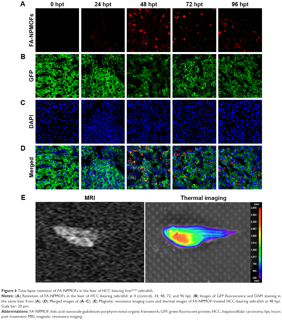
Fig. 6 Time-lapse retention of FA-NPMOFs in the liver of HCC-bearing krasG12V zebrafish. Notes: (A) Retention of FA-NPMOFs in the liver of HCC-bearing zebrafish at 0 (control), 24, 48, 72, and 96 hpt. (B) Images of GFP fluorescence and DAPI staining in the same liver from (A). (D) Merged images of (A–C). (E) Magnetic resonance imaging scans and thermal images of FA-NPMOF-treated HCC-bearing zebrafish at 48 hpt. Scale bar: 20 μm. Abbreviations: FA-NPMOF, folic acid-nanoscale gadolinium-porphyrin metal-organic framework; GFP, green fluorescent protein; HCC, hepatocellular carcinoma; hpt, hours post treatment; MRI, magnetic resonance imaging.