
Fig. 5
- ID
- ZDB-IMAGE-190403-7
- Publication
- Hsieh et al., 2018 - Global Expression Profiling Identifies a Novel Hyaluronan Synthases 2 Gene in the Pathogenesis of Lower Extremity Varicose Veins
- All Figures
- Figures for Hsieh et al., 2018
|
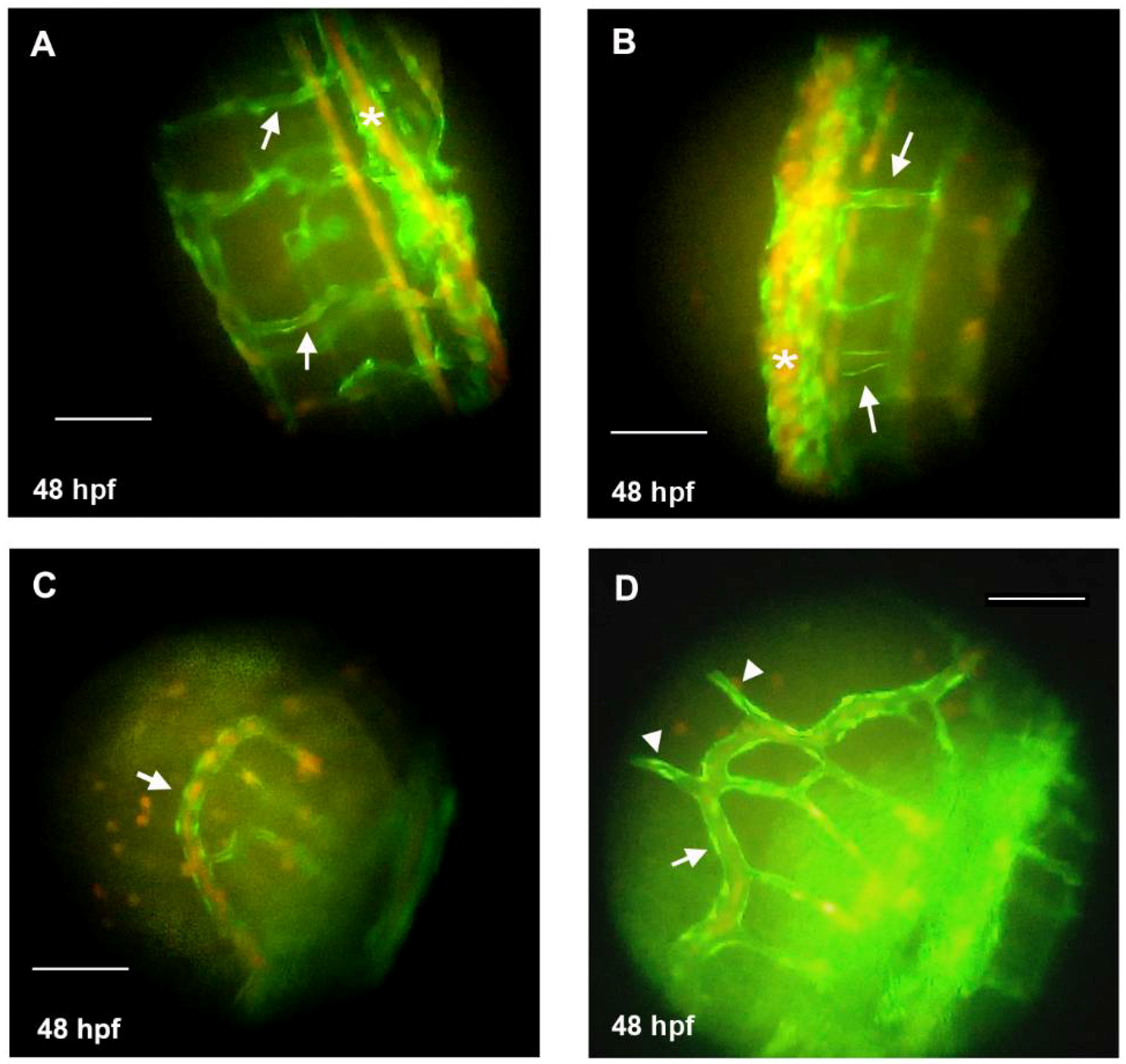
Fig. 5 Knockdown of hyaluronan synthases 2 gene (HAS2) in zebrafish embryos leads to dilated venous structure. Tg(fli1:egfp)xTg(gata1:dsRed) zebrafish with green fluorescence emitting vasculature are used to observe the anatomic change of vasculature. (A) Inter-segmental veins (ISVs) in the tail of a representative wild-type zebrafish (48 hpf) under fluorescence microscope (arrows); (B) Dilated ISVs in the tail of a representative HAS2-morpholino (MO) injected zebrafish (48 hpf) under fluorescence microscope (arrows); (C) The sub-intestinal vein (SIV) in the tail of a representative wild-type zebrafish (48 hpf) under fluorescence microscope (arrow); (D) The dilated SIV (arrow) with protruding branches (arrowheads) in the tail of a representative HAS2-morpholino (MO) injected zebrafish (48 hpf) under fluorescence microscope. Hpf indicates hours post-fertilization. Magnification is 400×. Scale bar = 100 μm.