
Fig. 5
- ID
- ZDB-IMAGE-190329-4
- Publication
- Pouchucq et al., 2018 - γ-Tubulin small complex formation is essential for early zebrafish embryogenesis
- All Figures
- Figures for Pouchucq et al., 2018
|
Fig. 5
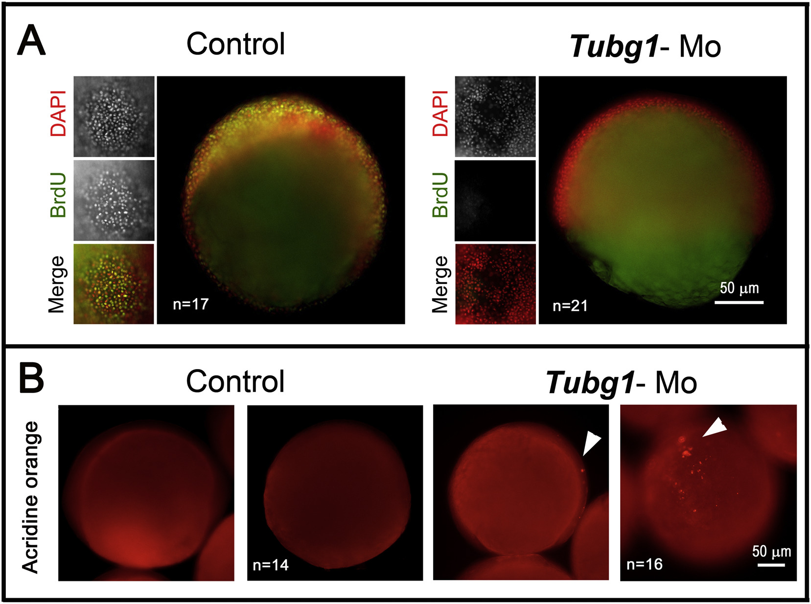
Cell proliferation arrest and apoptosis triggered in the absence of γ-TuSC formation. A. Zebrafish control and morphant embryos treated through the BrdU incorporation procedure. Embryos (8 hpf) were exposed to a BrdU pulse for 1 h, then fixed and treated for DNA labeling with DAPI (red) and immunolabeled for the incorporated BrdU (green). tubg1-Mo embryos did not incorporate BrdU in their DNA, indicating proliferation arrest. B. Apoptotic response revealed by TUNEL assay in embryos (8 hpf). The tubg1-morphant showed many clusters of cells positive for TUNEL stain (arrowheads) distributed throughout the blastoderm.
Reprinted from Mechanisms of Development, 154, Pouchucq, L., Undurraga, C.A., Fuentes, R., Cornejo, M., Allende, M.L., Monasterio, O., γ-Tubulin small complex formation is essential for early zebrafish embryogenesis, 145-152, Copyright (2018) with permission from Elsevier. Full text @ Mech. Dev.