
|
Fig. 4
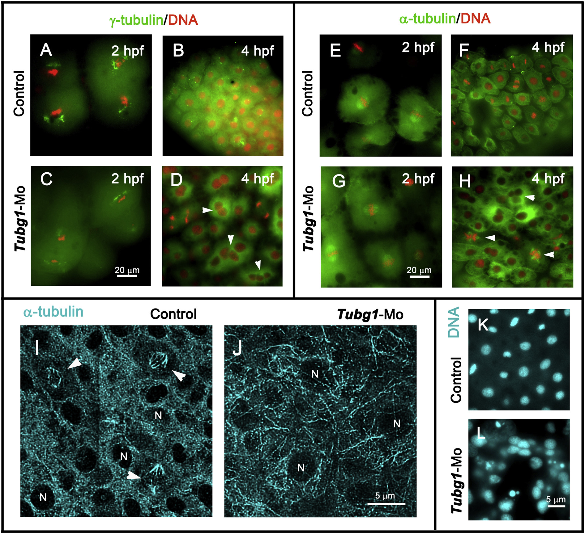
Nuclear and microtubule organization defects in γ-tubulin morphant embryos. A–D. Fluorescence microscopy images of control and morphant embryos at 2 (A and C) and 4 (B and D) hpf, showing centrosomal (γ-tubulin, green) and chromosomal (DAPI, red) distribution. E–H. anti-α-tubulin antibodies (green) and DAPI (red) double-stained embryos at 2 (E and G) and 4 (F and H) hpf. In the morphant embryos, γ-tubulin and microtubule organizations were severely affected and γ-TuSC formation impairment generated multinucleated blastomeres (arrowheads in D and H). I, J. Confocal projection images of α-tubulin-immunofluorescence staining of blastoderm in 8 hpf control and morphant embryos. Microtubule-stained spindle structures (arrowheads in I) and bundle organizations are shown. N, nucleus. K, L. DAPI-stained nuclei of control and tubg1-Mo embryos show a severe karyokinesis defect after γ-tubulin silencing.
Reprinted from Mechanisms of Development, 154, Pouchucq, L., Undurraga, C.A., Fuentes, R., Cornejo, M., Allende, M.L., Monasterio, O., γ-Tubulin small complex formation is essential for early zebrafish embryogenesis, 145-152, Copyright (2018) with permission from Elsevier. Full text @ Mech. Dev.