
Fig. 1
- ID
- ZDB-IMAGE-190329-1
- Genes
- Antibodies
- Publication
- Pouchucq et al., 2018 - γ-Tubulin small complex formation is essential for early zebrafish embryogenesis
- All Figures
- Figures for Pouchucq et al., 2018
|
Fig. 1
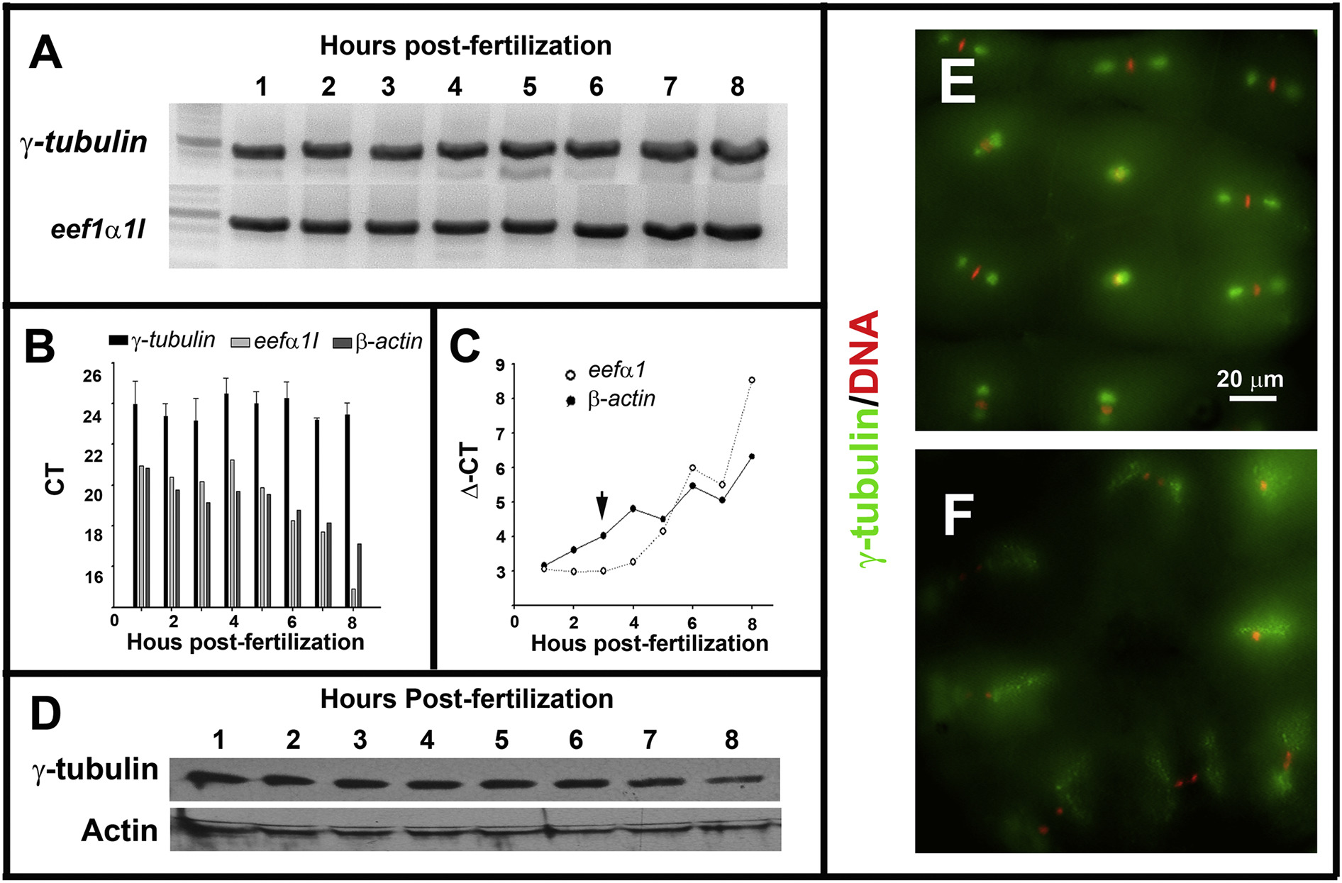
Maternal-zygotic zebrafish γ-tubulin expression and subcellular localization during early development. A. RT-PCR analysis revealed by electrophoresis in agarose gel showed that γ-tubulin mRNA is detected during the first 8 h of the zebrafish embryogenesis. B. qPCR analysis indicated that γ-tubulin mRNA expression levels were invariant during the first 8 h of zebrafish embryogenesis. β-actin and eef1a were used as reference genes. C. Immunoblot analysis showed that γ-tubulin protein is present during the cleavage (1–3 hpf) and post-MBT stages (3–8 hpf) of zebrafish development. Actin was used as a loading control. D–E. Fluorescence microscopy images of zebrafish embryos (2 hpf) double-stained with anti-γ-tubulin antibody (green) and DAPI (red). Metaphase (E), anaphase, and telophase (F) stages of the cell cycle are shown.
Reprinted from Mechanisms of Development, 154, Pouchucq, L., Undurraga, C.A., Fuentes, R., Cornejo, M., Allende, M.L., Monasterio, O., γ-Tubulin small complex formation is essential for early zebrafish embryogenesis, 145-152, Copyright (2018) with permission from Elsevier. Full text @ Mech. Dev.