
|
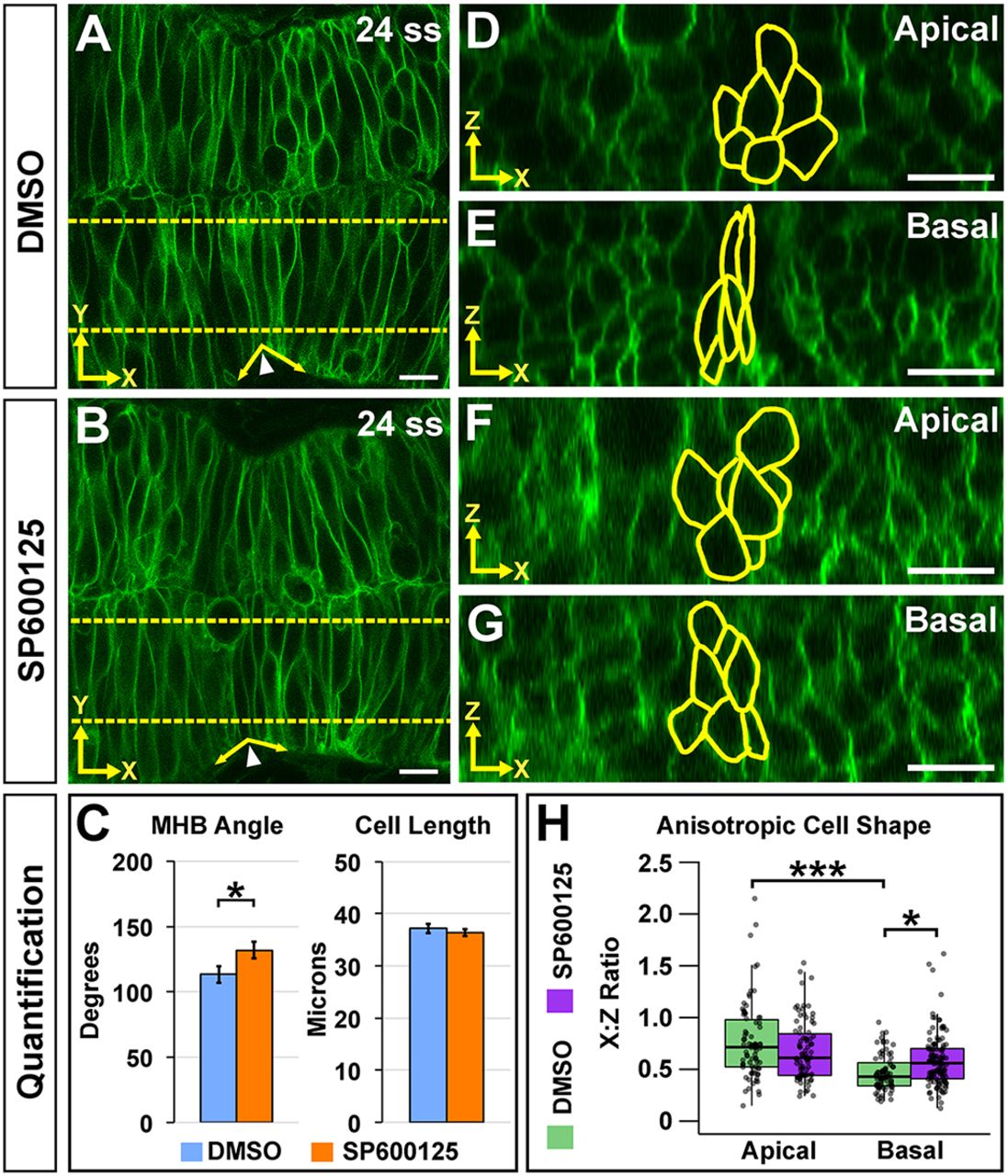
Fig. 7 JNK is required for basal anisotropic MHBC cell shape. (A,B) Live confocal imaging of 24 ss embryos injected with memGFP and treated with DMSO (A) or SP600125 (B). (C) Quantification and comparison of MHB angle and length. Data are represented as mean±s.e.m. of three independent experiments. (D,E) Apical (D) and basal (E) digital slices of DMSO-treated embryos at 24 ss. (F,G) Apical (F) and basal (G) digital slices of SP600125-treated embryos at 24 ss. MHBC cells are outlined in yellow in apical (D,F) and basal (E,G) digital slices. (H) Quantification of anisotropic cell shape using x:z ratio in DMSO- and SP600125-treated embryos. Boxplots indicate the 25th and 75th percentiles and the median. Three independent experiments are represented. DMSO, n=6; SP600125, n=6. *P<0.05, ***P<0.005. Arrowheads indicate MHBC and arrows indicate MHB tissue angle. Scale bars: 10 μm.