
|
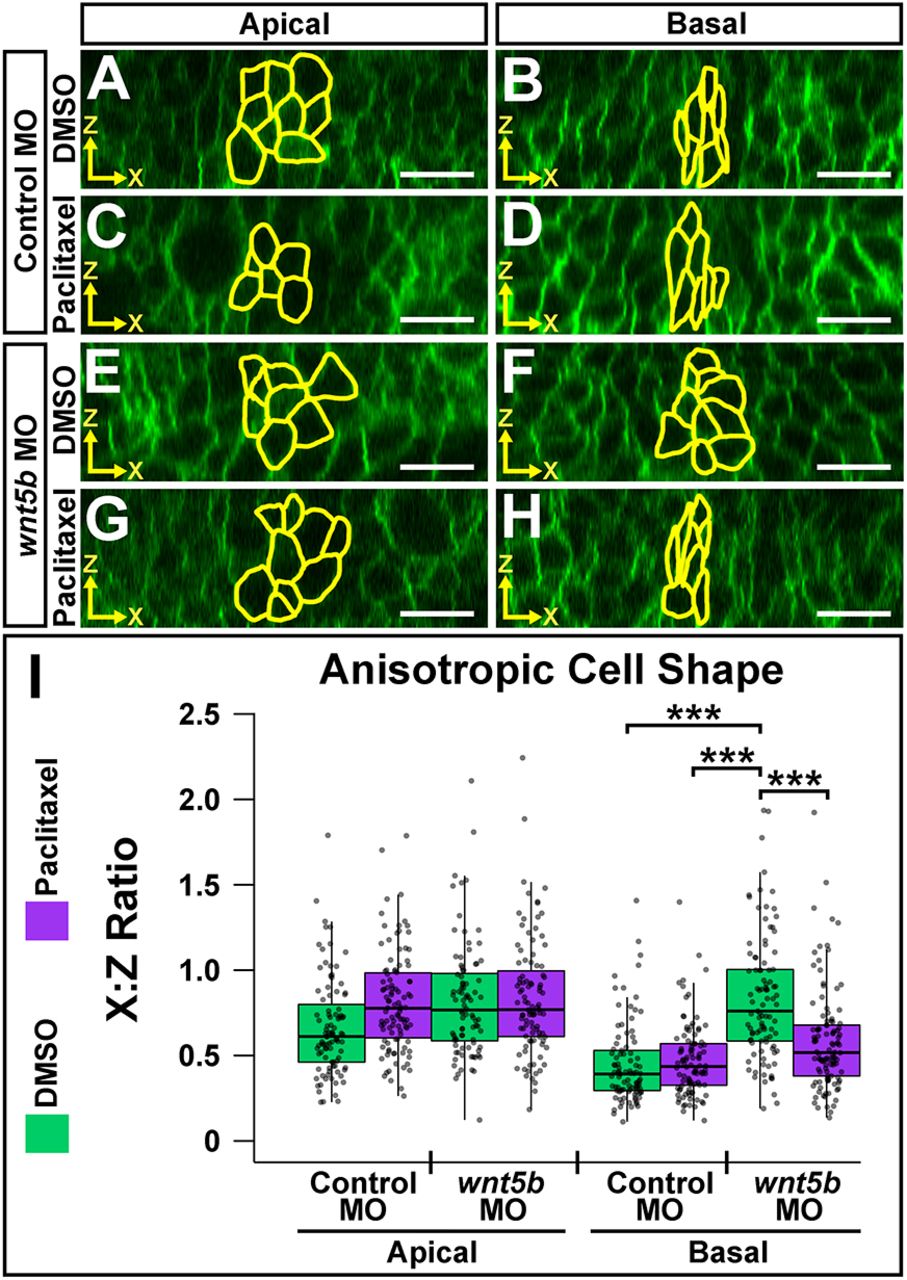
Fig. 5 Microtubule filament stability is required for Wnt5b-mediated basal anisotropic cell shape. (A-H) Digital slices from z-series images of wild-type embryos co-injected with memGFP and control MO (A-D) or wnt5b MO (E-H). Embryos were treated at 18 ss with DMSO (A,B,E,F) or paclitaxel (C,D,G,H) and imaged at 24 ss. MHBC cells are outlined in yellow in apical (A,C,E,G) and basal (B,D,F,H) digital slices. (I) Quantification of anisotropic cell shape using x:z ratio. Apical measurements were compared post-hoc with the control MO DMSO apical condition. Basal measurements were compared post-hoc with the wnt5b MO DMSO basal condition. Boxplots indicate the 25th and 75th percentiles and the median. Four independent experiments are represented. Control DMSO, n=7; control paclitaxel, n=9; wnt5b MO DMSO, n=8; wnt5b MO paclitaxel, n=8. ***P<0.005. Scale bars: 10 μm.