
|
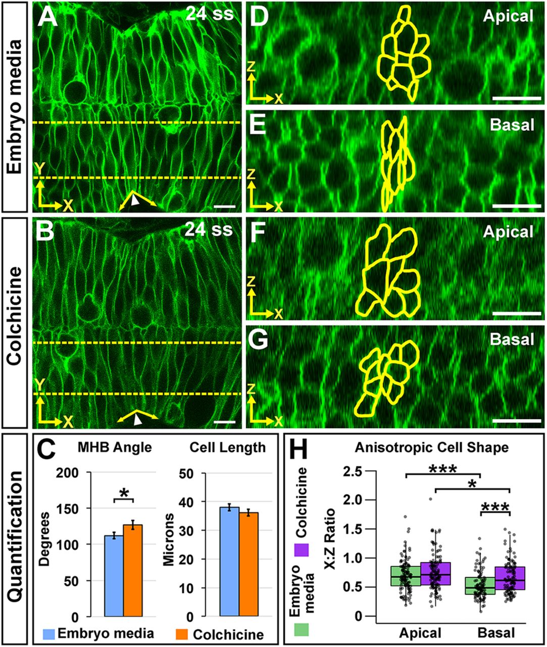
Fig. 4 Microtubule polymerization is required for basal anisotropic cell shape at the MHBC. (A,B) Live confocal images of 24 ss memGFP-injected wild-type embryos treated at 18 ss with embryo media (A) or colchicine (B). (C) Quantification of MHB tissue angle and MHBC cell length. Data are represented as mean±s.e.m. of three independent experiments. (D,E) Apical (D) and basal (E) digital slices of embryo media-treated embryos. (F,G) Apical (F) and basal (G) digital slices of colchicine-treated embryos. MHBC cells are outlined in yellow. (H) Quantification of anisotropic cell shape using x:z ratio. Boxplots indicate the 25th and 75th percentiles and the median. Three independent experiments are represented. Control embryo media, n=7; colchicine, n=8. *P<0.05, ***P<0.005. Arrowheads indicate MHBC and arrows indicate MHB tissue angle. Scale bars: 10 μm.