
|
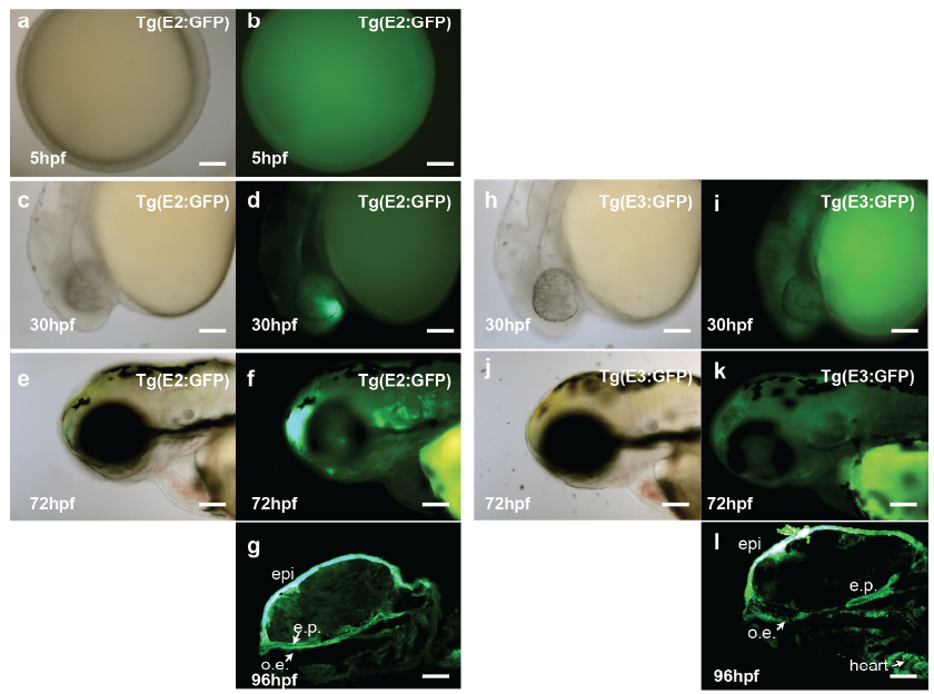
Fig. S2
Stable transgenic zebrafish lines showing in vivo enhancer activities patterns of E2 and E3 in different development stages.
In 5hpf, F2 embryos of Tg(E2:GFP) exhibit universal enhancer activity (a, b). In 30 hpf, F2 embryos of Tg(E2:GFP) exhibit GFP activity in the forebrain (c, d), and F2 embryos of Tg(E3:GFP) exhibit GFP activity in central nervous system (h, i). In 72 hpf, F2 embryos of Tg(E2:GFP) exhibit GFP activity in lower jaw, otic vesicle, head epidermis and forebrain (e, f), and F2 embryos of Tg(E3:GFP) exhibit GFP activity in lower jaw and head epidermis (j, k). In 96hpf, sagittal section of F2 embryos of Tg(E2:GFP) (g) showing in vivo activity of E2 in oral epithelium (o.e.), ethmoid plate (e.p.) and head epidermis (epi), and sagittal section of F2 embryos of Tg(E3:GFP) (l) showing in vivo activity of E3 in oral epithelium, ethmoid plate, head epidermis and heart. Scale bar =100μm.