
Fig. 4
- ID
- ZDB-IMAGE-190118-4
- Publication
- Gutzman et al., 2018 - Basal constriction during midbrain-hindbrain boundary morphogenesis is mediated by Wnt5b and focal adhesion kinase
- All Figures
- Figures for Gutzman et al., 2018
|
Fig. 4
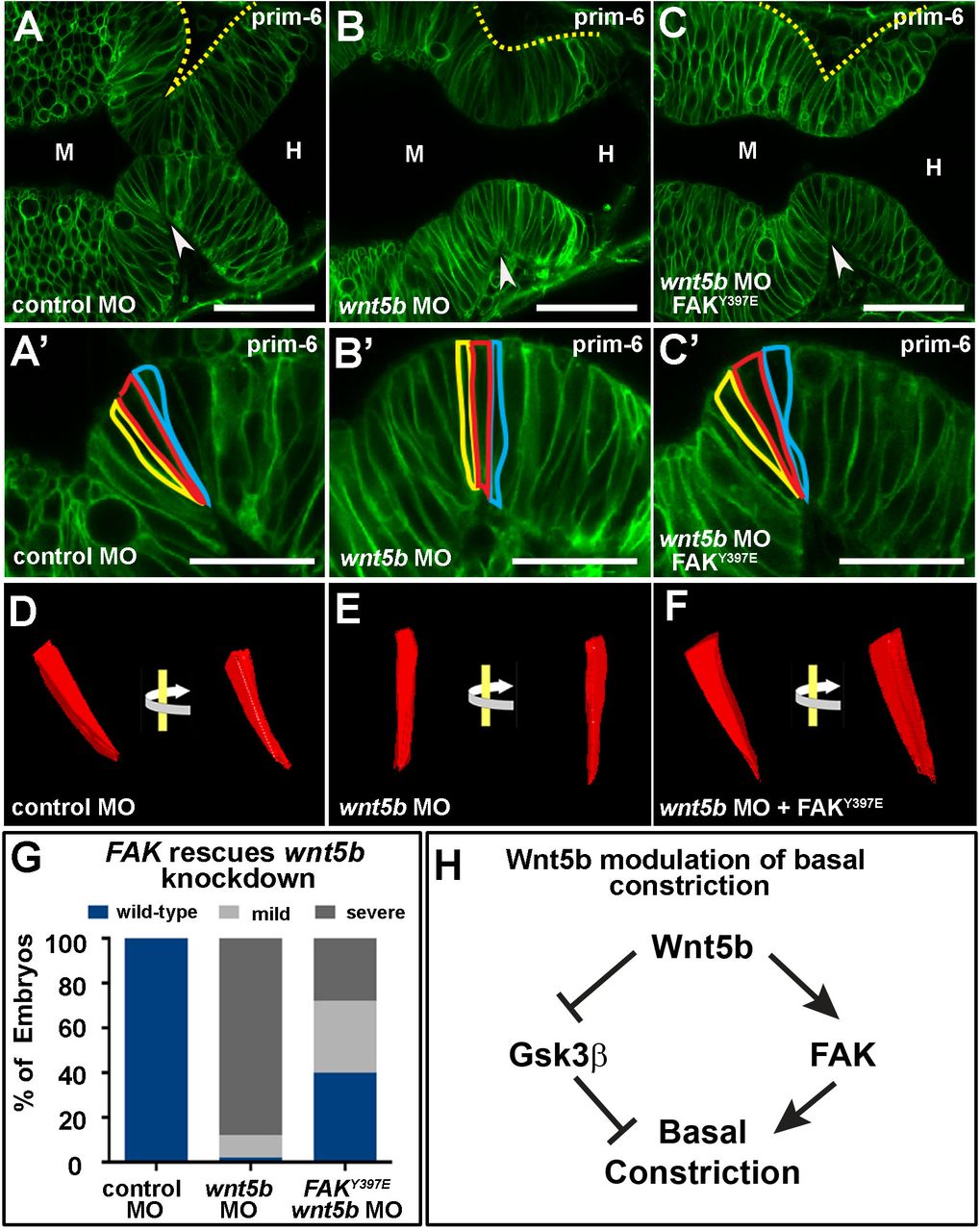
Fak rescues effects of wnt5b knockdown on basal constriction. (A–C′) Live confocal images of the MHB region in prim-6 stage embryos. Single-cell wild-type embryos were co-injected with mGFP and control MO (A,A′), wnt5b MO (B,B′), or wnt5b MO and FAKY397E mRNA (C,C′). (D–F) 3D reconstruction of cells outlined in (A′–C′) using 3D doctor with view of the same cell rotated 45°. (G) Quantification of MHBC gross morphology and basal constriction following FAK397E mRNA rescue of wnt5b knockdown (n>60 per condition). (H) Model pathway for Wnt5b regulation of basal constriction. Arrowheads indicate MHBC. M, midbrain. H, hindbrain. Scale bars: A–C, 50 µm; A′–C′, 25 µm.