
Fig. S3
- ID
- ZDB-IMAGE-190108-10
- Publication
- Cantù et al., 2018 - Mutations in Bcl9 and Pygo genes cause congenital heart defects by tissue-specific perturbation of Wnt/β-catenin signaling.
- All Figures
- Figures for Cantù et al., 2018
|
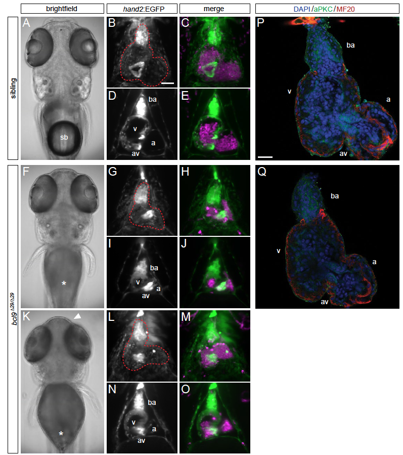
Fig. S3
Cardiac phenotypes of zebrafish bcl9Δ29 mutants.
(A-O) SPIM images of hand2:EGFP;drl:mCherry-expressing wildtype siblings and homozygous bcl9Δ29 embryos at 5 dpf; ventral views, anterior to the top, imaged after viable heart-stopping BDM treatment, two individual bcl9Δ29-mutant embryos are shown. A,F,K, bright field views to illustrate the absent swimbladder in mutants (sb, asterisks in F,K) and different expressivity of craniofacial defects (arrowhead, K); B-O maximum-intensity projection fluorescence close-ups of the heart (red dotted outlines in B,G,L); D,E,I,J,N,O depict optical sections at AV canal level. (P,Q) Confocal sections of sibling control (P) and homozygous bcl9Δ29 (Q) hearts, dissected and stained at 5 dpf to reveal the details of cardiac architecture. Also compare to Figure 2. ba, bulbus arteriosus; a, atrium; v ventricle; av, atrioventricular canal. Scale bars B-E,G-J,L-O 100 m, S,T 20 m.