
Fig. 1
- ID
- ZDB-IMAGE-181206-13
- Genes
- Publication
- Xu et al., 2018 - Inhibition of TBK1/IKKε Promotes Regeneration of Pancreatic β-cells
- All Figures
- Figures for Xu et al., 2018
|
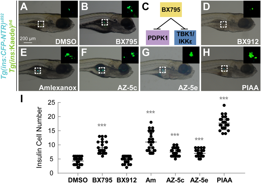
Fig. 1
TBK1/IKKε inhibition augments β-cell regeneration in a zebrafish model of type 1 diabetes. (A, B, D–H) Bright-field images combined with fluorescent images showing the overall morphology and [Tg(ins:CFP-NTR)s892; Tg(ins:Kaede)jh6] expression (green) of larvae at 48 hpa treated with DMSO (A), BX795 (B), BX912 (D), amlexanox (E), AZ-5c (F), AZ-5e (G), and PIAA (H), respectively. While TBK1/IKKε-Is substantially expanded [Tg(ins:CFP-NTR)s892; Tg(ins:Kaede)jh6]-expressing cell population (white squares and insets) during regeneration (B and E–H) compared to DMSO (A), the PDPK1 inhibitor BX912 showed minimum effect (D). (C) BX795 is a dual inhibitor of PDPK1 and TBK1/IKKε. (I) Quantification of the number (mean ± SD) of total regenerated β-cells at 48 hpa (in A-B and D-H; 4.3 ± 1.3 (DMSO), 9.0 ± 2.2 (BX795), 4.7 ± 1.2 (BX912), 11.0 ± 3.4 (amlexanox), 7.4 ± 1.4 (AZ-5c), 7.0 ± 1.3 (AZ-5e), and 17.7 ± 2.4 (PIAA)). Cells in 20 planes of confocal images from 25 individual larvae were counted per condition. ***P < 0.001.