
Image
|
Figure Caption
Fig. S3
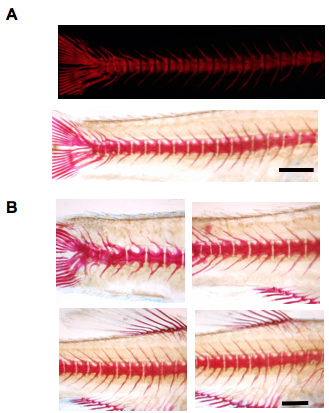
Additional Images of abnormal slc39a8 KO zebrafish after alizarin red staining at 12 weeks post-fertilization. (A) A single mutant slc39a8 zebrafish demonstrating spinal fusion by fluorescent or light microscopy. (B) Various skeletal abnormalities were observed among slc39a8 KO zebrafish including fused vertebrae and abnormal bone growth along the spine (4 different fish shown). Shown are homozygous mutant slc39a8 zebrafish produced from a cross between two heterozygous mutant slc39a8 zebrafish and genotyped for the mutation. Scale bar 2mm.
Figure Data
Acknowledgments
This image is the copyrighted work of the attributed author or publisher, and
ZFIN has permission only to display this image to its users.
Additional permissions should be obtained from the applicable author or publisher of the image.
Full text @ Nat. Commun.