
Image
|
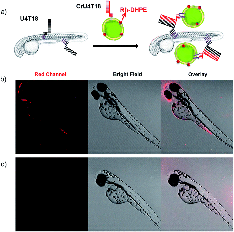
Figure Caption
Fig. 2
DNA duplex formation between U4T18 and CrU4T18 decorated liposomes on the surface of zebrafish. (a) Schematic representation of liposomes docking on the surface of zebrafish embryos by lipid–DNA hybridization. Confocal images of 2 dpf zebrafish treated with (b) U4T18 for 1 h, followed by incubation with CrU4T18 decorated liposomes or (c) treated with 1 μM CrU4T18 decorated liposomes in absence of U4T18. The concentration of total lipids (DOPC![[thin space (1/6-em)]](https://www.rsc.org/images/entities/char_2009.gif) :DOPE:Chol = 2:1:1 mol%) was 0.5 mM. Red channel: Rh-DHPE.
:DOPE:Chol = 2:1:1 mol%) was 0.5 mM. Red channel: Rh-DHPE.
Acknowledgments
This image is the copyrighted work of the attributed author or publisher, and
ZFIN has permission only to display this image to its users.
Additional permissions should be obtained from the applicable author or publisher of the image.
Full text @ Chem Sci