
Fig. 5
- ID
- ZDB-IMAGE-181114-6
- Publication
- Sun et al., 2018 - Maternal Ybx1 safeguards zebrafish oocyte maturation and maternal-to-zygotic transition by repressing global translation
- All Figures
- Figures for Sun et al., 2018
|
Fig. 5
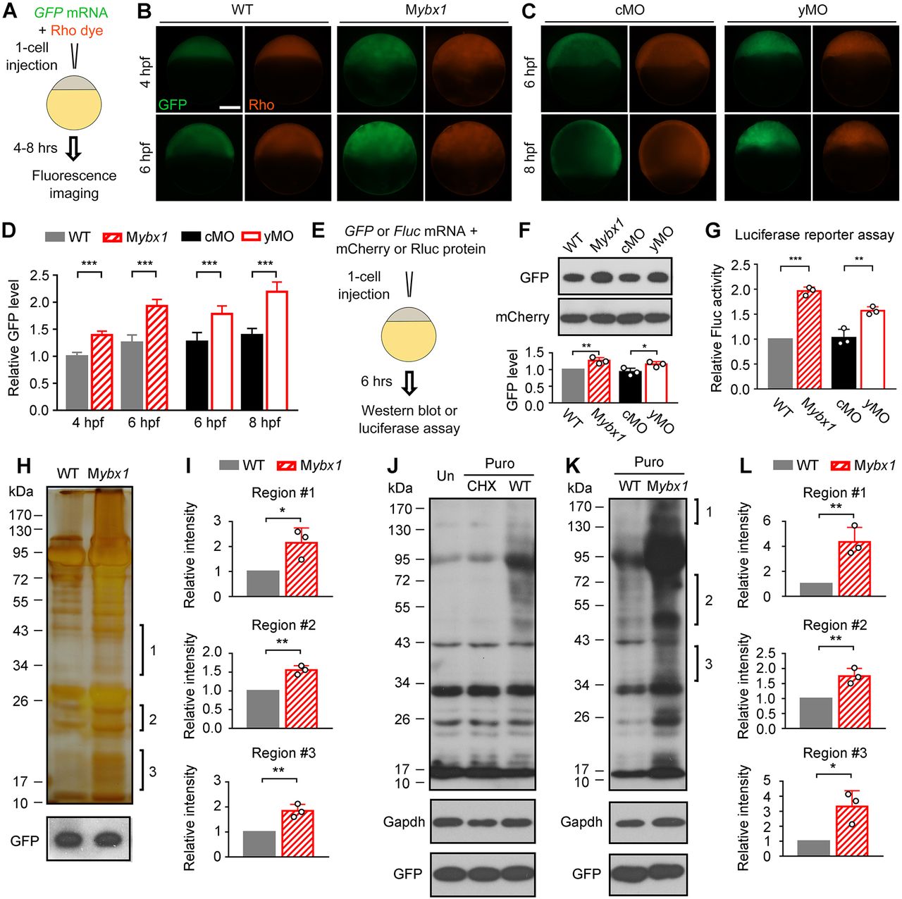
Loss of maternal Ybx1 elevates global translation level. (A) Schematic of the fluorescent reporter translation assay. (B,C) Fluorescent images showing GFP reporter levels with rhodamine (Rho) control levels in WT, Mybx1, cMO- and yMO-injected live embryos at indicated stages. Scale bar: 200 μm. (D) Measurement of GFP reporter intensities relative to Rho. (E) Schematic of reporter translation assays using western blotting and luciferase assay. (F) Western blotting analysis of GFP reporter levels at 6 hpf. (G) Measurement of Fluc activities relative to Rluc. (H) SDS-PAGE and silver staining showing global protein levels in embryos. Three regions for measurement are indicated. 3 hpf embryos were lysed for the SDS-PAGE. GFP protein was injected at the one-cell stage as the loading control. (I) Quantification of relative signal intensities of three regions from H. (J) Western blot for the puromycin incorporation assay. GFP protein was injected as the loading control. Gapdh levels are also shown. Un, untreated; Puro, puromycin; CHX, cycloheximide. (K) Puromycin incorporation levels in WT and Mybx1 embryos shown by western blotting. The injected GFP protein served as the loading control. (L) Relative puromycin signal intensities of three regions from western blots in K. *P<0.05, **P<0.01, ***P<0.001; ns, not significant; n=8 embryos for D, n=3 biological replicates for F,G,I,L; Student's t-test.