
|
Fig. S7
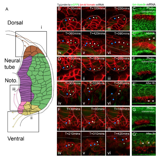
Niches of progenitor cells with non-muscle cell fates and the corresponding migratory paths, related to Figure 5 and 7
(A) Schematic diagram of niches of progenitor cells with non-muscle cell fates and corresponding migratory routes. Three niches revealed in this study locate at dorsal margin (i), lateral ventral margin (ii) and medial ventral margin (iii) of somite. (B-G’) Cell tracking reveals the niches and corresponding migratory routes. (B-C) Progenitor cells stemmed from dorsal margin (i) migrate dorsally and move into the interface between myotome and neural tube. (D-E) Progenitor cells stemmed from lateral ventral margin (ii) migrate ventrally. (F-G) Progenitor cells stemmed from medial ventral margin (iii) migrate to the interface between notochord and myotome. (B, D, F) Cell tracking based on time-lapse imaging. White lines denote to the boundary of myotome. (C, E, G) Cell tracking based on photo-conversion of Kaede at the dorsal and ventral margin. White short arrows label the corresponding non-muscle progenitors at 8 hours after photo-conversion.
Reprinted from Developmental Cell, 46, Yin, J., Lee, R., Ono, Y., Ingham, P.W., Saunders, T.E., Spatiotemporal Coordination of FGF and Shh Signaling Underlies the Specification of Myoblasts in the Zebrafish Embryo, 735-750.e4, Copyright (2018) with permission from Elsevier. Full text @ Dev. Cell