
Fig. S3
- ID
- ZDB-IMAGE-181018-13
- Publication
- Du et al., 2018 - A transgenic zebrafish model for in vivo long-term imaging of retinotectal synaptogenesis
- All Figures
- Figures for Du et al., 2018
|
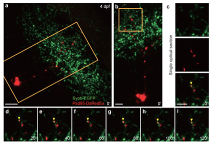
Fig. S3
Example of simultaneous imaging of pre- and postsynaptic sites of retinotectal synapses.
(a) First image of a time-lapse series showing Psd95-DsRedEx labelled postsynaptic sites in the dendritic arbor of a tectal neuron (red) and the Sypb-EGFP labelled presynaptic terminals on the axonal arbor of multiple RGCs (green) in a 4-dpf PGUSG larva with transient expression of elavl3:psd95-DsRedEx. The nasal is upwards. Scale bar, 10 μm. (b) Enlarged view of the boxed region in (a). Scale bar, 5 μm. (c) Enlarged single optical section of the boxed region in (b) showing single-channel images and the composite image. The yellow arrowhead (bottom) indicates the association of a presynaptic terminal labelled by Sypb-EGFP (green, top) with a postsynaptic site labelled by Psd95-DsRedEx (red, middle). Scale bar, 5 μm. (c-i) 2-h time series at 20 min intervals showing the long-term stable overlap of pre- and postsynaptic labels (yellow arrowhead). Single optical section of the same area as in (c) was shown. Time in minutes is indicated in the bottom right corner of each panel.