
Image
|
Figure Caption
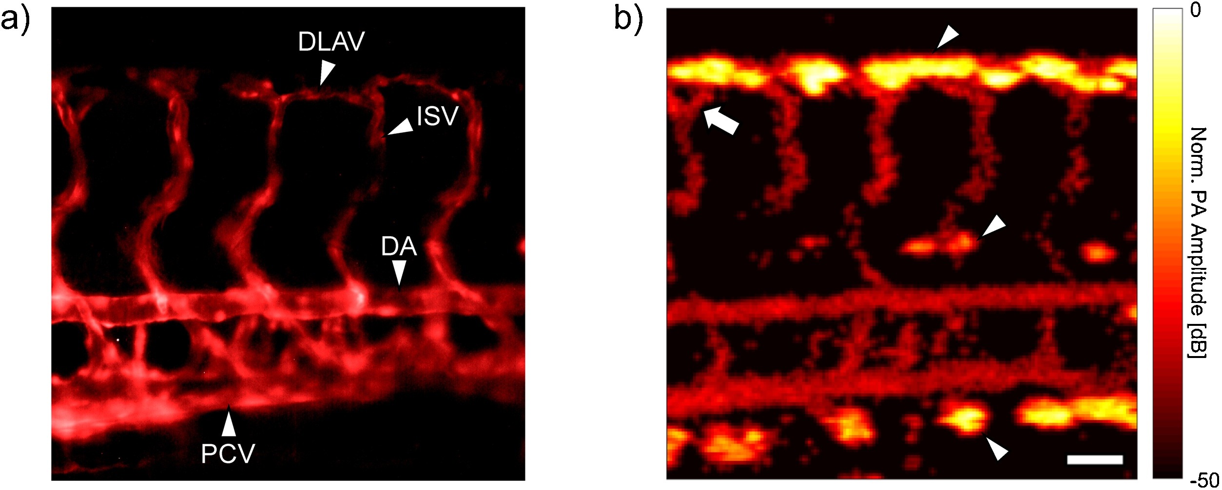
Fig. 2
a) Fluorescence image of the GFP expressing endothelial cells in the trunk of a Tg (flk1:GFP) larval zebrafish. b) PA image of the same fish. Erythrocytes within the lumenized vessels allow for visualization of perfused vasculature. The arrow denotes a bifurcation in one of the ISVs, while the arrowheads indicate melanin spots. The scale bar is 50 μm and can be applied to both images. DLAV = Dorsal longitudinal anastomotic vessel, ISV = Intersegmental vessel, DA = Dorsal aorta, PCV = Posterior cardinal vein.
Acknowledgments
This image is the copyrighted work of the attributed author or publisher, and
ZFIN has permission only to display this image to its users.
Additional permissions should be obtained from the applicable author or publisher of the image.
Full text @ Photoacoustics