
IMAGE
Fig. S1
- ID
- ZDB-IMAGE-181004-6
- Publication
- Wu et al., 2018 - Il34-Csf1r Pathway Regulates the Migration and Colonization of Microglial Precursors
- All Figures
- Figures for Wu et al., 2018
Image
|
Figure Caption
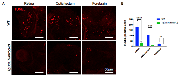
Fig. S1
Tg(Xla.Tubb:bcl-2) Transgene Successfully Blocks Neuronal Apoptosis in Most CNS Regions. Related to Figure 1.
(A and B) Whole mount Terminal deoxynucleotidyl transferase dUTP nick end labeling (TUNEL) assay followed by transverse section showing representative images of apoptotic cells (red) (A) and quantification (B) of apoptotic cells in different CNS regions of 3 dpf WT or Tg(Xla.Tubb:bcl-2) embryos injected with pu.1 morpholino. n = 4 and 3 for WT and Tg(Xla.Tubb:bcl-2) embryos, respectively. Values represent means with SD. ns, P>0.05; ***, P ≤ 0.001; ****, P ≤ 0.0001.
Figure Data
Acknowledgments
This image is the copyrighted work of the attributed author or publisher, and
ZFIN has permission only to display this image to its users.
Additional permissions should be obtained from the applicable author or publisher of the image.
Reprinted from Developmental Cell, 46, Wu, S., Xue, R., Hassan, S., Nguyen, T.M.L., Wang, T., Pan, H., Xu, J., Liu, Q., Zhang, W., Wen, Z., Il34-Csf1r Pathway Regulates the Migration and Colonization of Microglial Precursors, 552-563.e4, Copyright (2018) with permission from Elsevier. Full text @ Dev. Cell