
Image
|
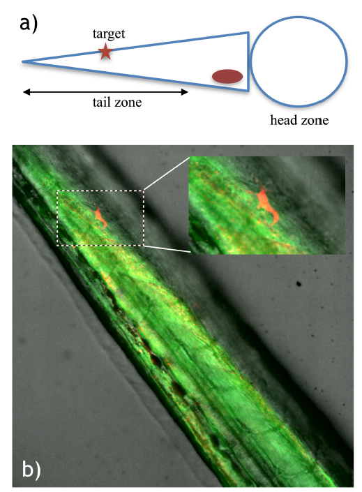
Figure Caption
Fig. S1
Single cell labeling in the zebrafish tail. a) Diagram depicting the embryo positioning for the experiment and the target area in the embryo tail. b) Labeled cell identified by red fluorescence (mCherry expression), demonstrating successful photoactivation in the zebrafish tail. The inset shows a magnification of the boxed area.
Acknowledgments
This image is the copyrighted work of the attributed author or publisher, and
ZFIN has permission only to display this image to its users.
Additional permissions should be obtained from the applicable author or publisher of the image.
Full text @ Light Sci Appl