
Image
|
Figure Caption
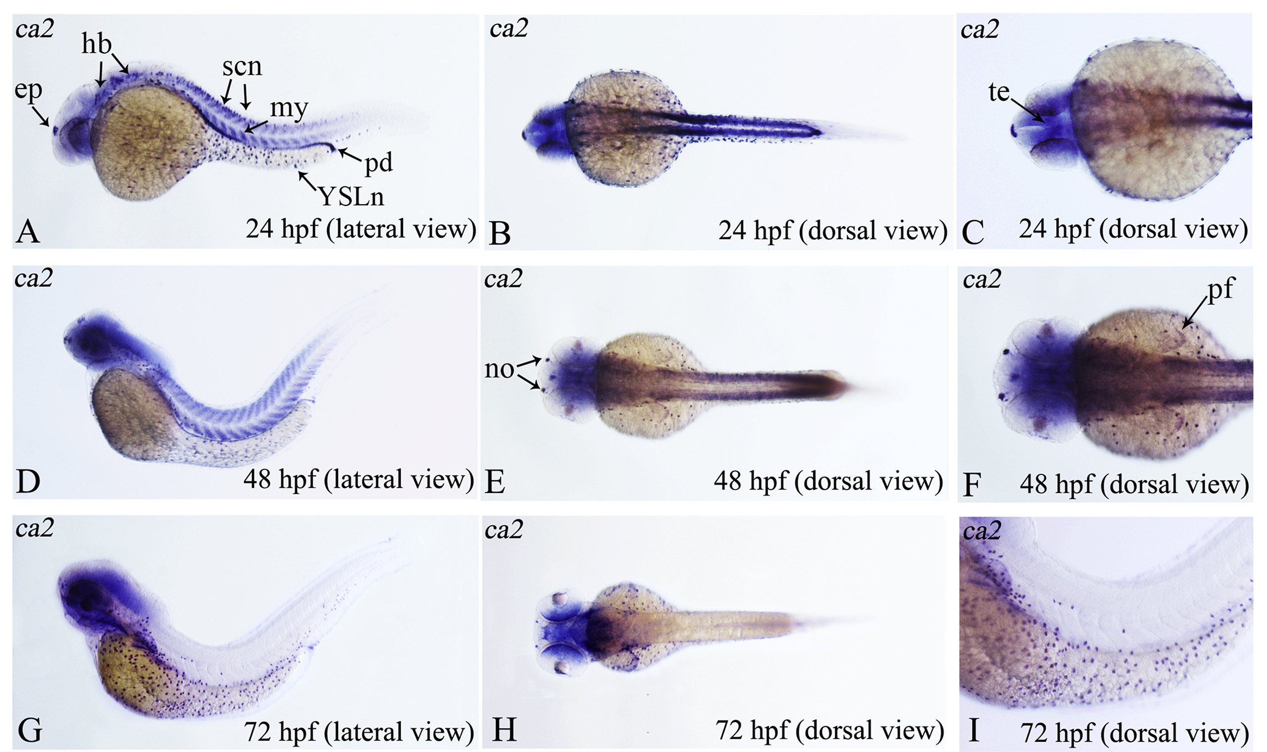
Fig. 8
The expression pattern of ca2 detected by WISH. Stages of embryonic development: A-C. 24 hpf; D-F. 48 hpf; G-I. 72 hpf (hpf, hours post-fertilization); C, F, I. High magnification views of B, E and G respectively. ep: epiphysis; hb: hindbrain; my: myotomes; no: nose; pd: pronephric ducts; pf: pectoral fin; scn: spinal cord neurons; te: tegmentum; YSLn: YSL nuclei.
Figure Data
Acknowledgments
This image is the copyrighted work of the attributed author or publisher, and
ZFIN has permission only to display this image to its users.
Additional permissions should be obtained from the applicable author or publisher of the image.
Reprinted from Gene expression patterns : GEP, 29, Feng, S., Wang, S., Wang, Y., Yang, Q., Wang, D., Li, H., Identification and expression of carbonic anhydrase 2, myosin regulatory light chain 2 and selenium-binding protein 1 in zebrafish Danio rerio: Implication for age-related biomarkers, 47-58, Copyright (2018) with permission from Elsevier. Full text @ Gene Expr. Patterns