
|
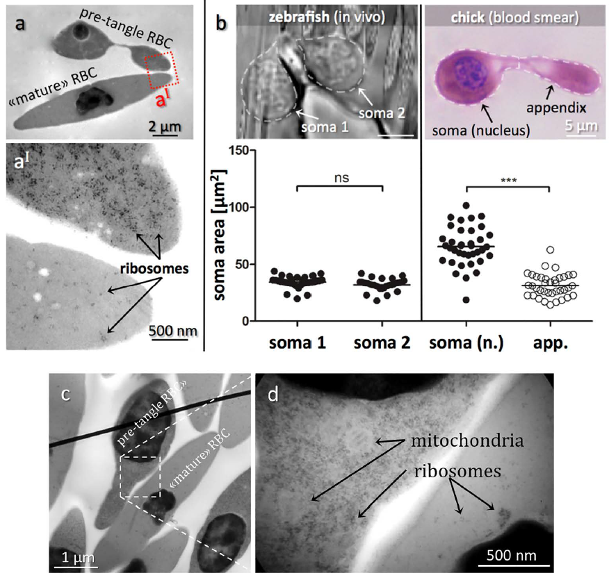
Fig. S1
Transmission electron micrographs of RBCs in blood of fish and chick embryos.
(a) Electron micrographs comparing pre-tangle and mature chick RBCs. (a´) Zoomed region indicated in (a) in a pre-tangle RBC (top) contains abundant free ribosomes in contrast to the mature RBC (bottom). Other side-by-side comparisons of pre-tangling versus mature chick RBCs as shown in (c + d) confirmed the far higher quantity of ribosomes and mitochondria in immature RBCs relative to fully differentiated cells. (b) Comparing somata sizes of dumbbellshaped zebrafish and chick RBCs. In the zebrafish, both somata are nucleated and of similar size. In contrast, only one soma is nucleated in the chick RBC, which is significantly larger in size than the other soma. ***P<0.001.