
|
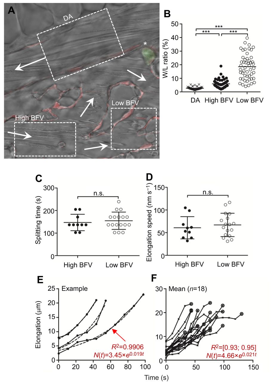
Fig. 2
RBC splitting is independent of the incident blood flow velocity in zebrafish embryos in vivo. To assess the impact of blood flow velocity (BFV) as a major shear force determinant on splitting time and elongation speed during RBC splitting, we compared RBC splitting at two regions receiving either high or low BFV. (A) Three regions in a Tg(fli1a:RedX;mpx:eGFP) fish, the dorsal aorta (DA), high BFV and low BFV, are indicated. Arrows mark the respective blood flow direction. The asterisk indicates a primitive neutrophil. (B) BFV was deduced by the width/length (W/L) ratio of adjacent circulating blood cells. Images were acquired throughout a constant period of time. In regions with high BFV (i.e. DA), RBCs are elongated (i.e. L>W, low W/L ratio), while at low BFV, RBCs appear more rounded (i.e. increased W/L ratio). Therefore, the W/L ratio serves as a proxy for the local BFV. ***P<0.001. (C) Splitting time and (D) elongation speed were both independent on BFV. (E,F) Exponential growth (E) and elongation length (F). Once RBCs are tangled at vascular structures (phase ii), the soma-to-soma connection subsequently elongates (phase iii). The process of elongation can be mathematically expressed using exponential growth equations. This implies that the longer a cell is tangled, the faster its retained connection elongates. On average, elongation was represented by the equation N(t)=4.66×e0.021t with a regression coefficient (R2) between 0.93 and 0.95 (n=1 animal, n=18 events).