
|
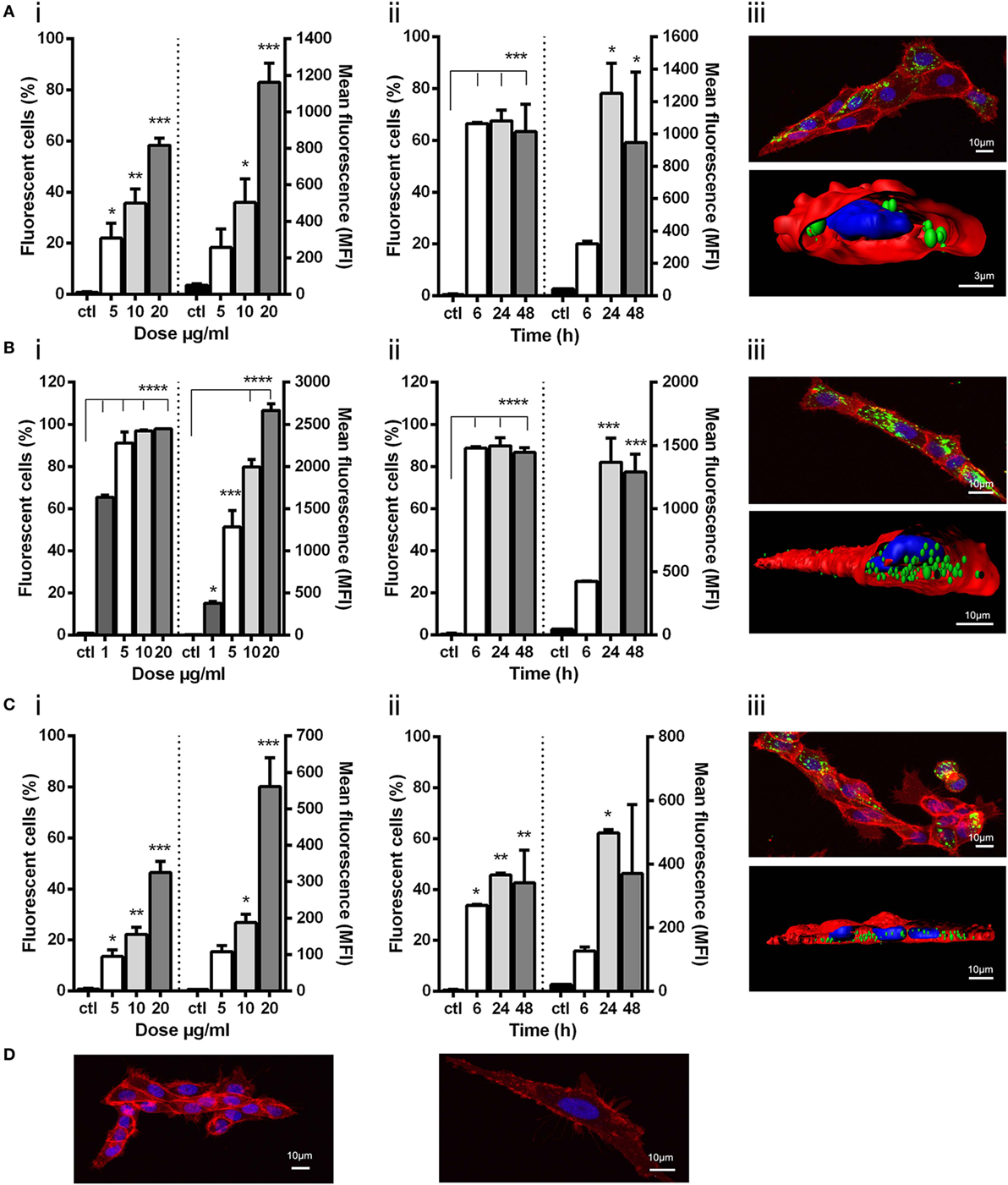
Fig. 2
Uptake of viral nanopellets (NPs) by ZFL. Fluorescently labeled NPs (A) IPNV-VP2NP, (B) VHSV-G-frg16NP, and (C) VNNV-CNP were added to ZFL. Control (ctl) was ZFL without NPs. (i) Dose–response. Cells incubated for 12 h with NPs (A,C) at 5–20 µg/ml, and (B) at 1–20 µg/ml in duplicate. (ii) Time course. NPs added to cells at 10 µg/ml (A,C), and 5 µg/ml (B) in duplicate and incubated for 6–48 h. Differences between means were analyzed by a one-way ANOVA with Dunnett’s multiple comparisons test, treatments versus control. Significance levels *p < 0.05; **p < 0.01; ***p < 0.001; ****p < 0.0001. (iii) Confocal microscopy and digitalized image (z-stacks) of ZFL cells after 14 h incubation with 20 µg/ml (A,C) and 10 µg/ml (B). NPs are green, cell membrane red, and nuclei blue. Control confocal image ZFL without NPs (D).