
|
Fig. 5
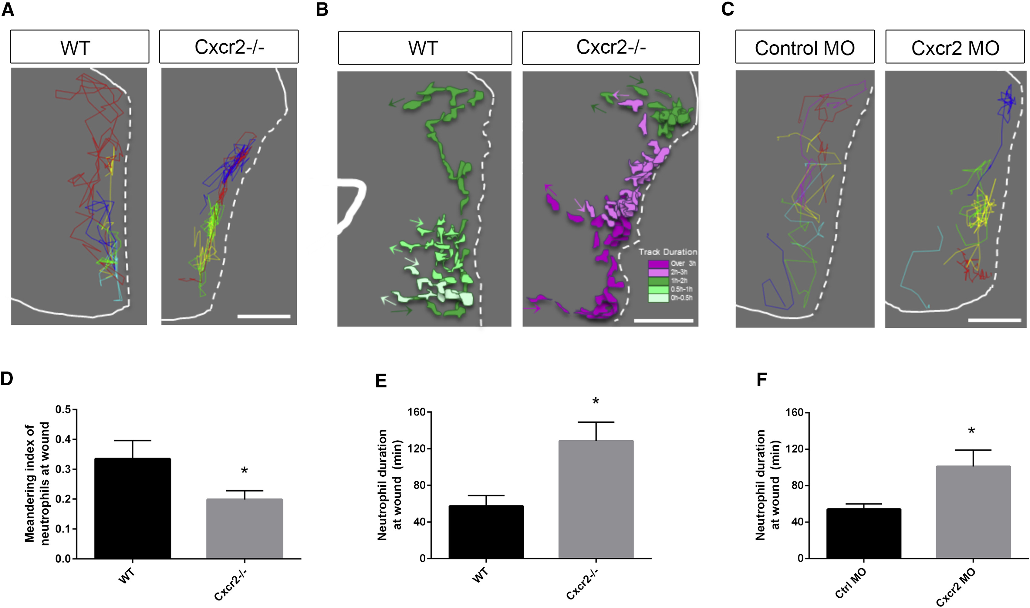
Interstitial Behavior in the Wound Microenvironment Is Altered in cxcr2-Deficient Zebrafish
(A) Representative image of neutrophil tracks within the tail fin of WT or Cxcr2−/− larvae. Each color track represents an individual neutrophil (white dotted line traces wound edge).
(B) Representative tracks of neutrophil morphology at the wound in WT or Cxcr2−/− larvae. Individual neutrophils are color coded to represent duration at the wound (see legend), with each colored track representing one neutrophil over the duration of its time at the wound.
(C) Representative neutrophil tracks at the wound of WT or cxcr2 morphant larvae.
(D) Average meandering index of neutrophils at the wound in WT is greater than in Cxcr2−/− larvae (n = 20, each).
(E and F) Average neutrophil duration at the wound is greater (E) in Cxcr2−/− compared to WT and (F) in cxcr2 MO compared to WT (n = 20, each). ∗p < 0.05.
Error bars represent SE.