
|
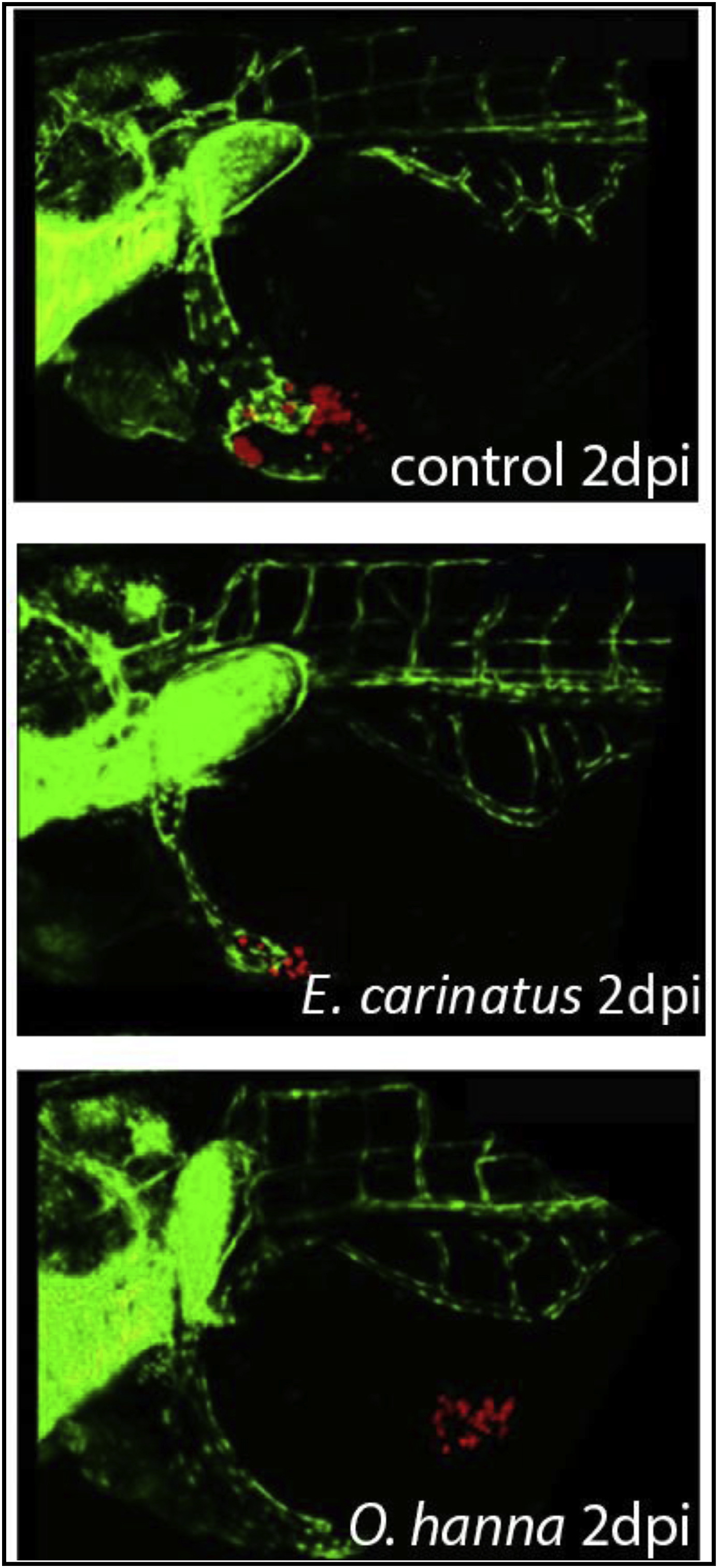
Fig. 7
Anti-angiogenic properties of O. hannah venom and E. Carinatus venom at 2 dpi. The figure shows three confocal micrographs of PaTu 8988t cells (red) transplanted into 48 hpf fli zebrafish embryos (green). The cells were dispersed in Matrigel, as a result of which they remain in the yolk sac. When O. hannah venom was added to the cells, inhibition of angiogenesis was observed. However, treatment of the fish with E. carinatus venom resulted in angiogenesis, like the controls. 20× magnification. (n = 20 embryos per tested snake venom). (For interpretation of the references to color in this figure legend, the reader is referred to the Web version of this article.)